


From Lipid Peroxidation to Patient Stratification in Ovarian Cancer
1Department of Oncology, Wuxi Maternity and Child Health Care Hospital, Women’s Hospital of Jiangnan University, Jiangnan University, Wuxi 214002, China
2Department of Oncology, Wuxi Maternal and Child Health Care Hospital, Wuxi Medical Center, Nanjing Medical University, Wuxi 214002, China
aThese authors contributed equally to this work.
*Correspondence to: Shaojie Zhao, Department of Oncology, Wuxi Maternity and Child Health Care Hospital, Women’s Hospital of Jiangnan University, Jiangnan University, Wuxi 214002, China, E-mail: zsjie2005@163.com; Yan Zhang, Department of Oncology, Wuxi Maternity and Child Health Care Hospital, Women’s Hospital of Jiangnan University, Jiangnan University, Wuxi 214002, China, E-mail: fuyou2007@126.com
Received: June 30 2026; Revised: August 16 2026; Accepted: September 17 2026; Published Online: October 23 2026
Cite this paper:
Yang X, Wang Y, You H et al. From Lipid Peroxidation to Patient Stratification in Ovarian Cancer. BIO Integration 2026; 7: 1–7.
DOI: 10.15212/bioi-2026-0128. Available at: https://bio-integration.org/
Download citation
© 2026 The Authors. This is an open access article distributed under the terms of the Creative Commons Attribution License (https://creativecommons.org/licenses/by/4.0/). See https://bio-integration.org/copyright-and-permissions/
Abstract
Ovarian cancer cells exploit the lipid-rich peritoneal microenvironment to sustain survival and dissemination. An increase in fatty acid uptake enriches the microenvironment in oxidizable membrane phospholipids and raises the risk of lipid peroxidation (LP)-driven ferroptosis. We argue that ovarian cancers exposed to chronic oxidative stress may acquire an enhanced dependence on ferroptosis defense pathways, particularly the solute carrier family 7 member 11 (SLC7A11)–glutathione (GSH)–glutathione peroxidase 4 (GPX4) axis, the ferroptosis suppressor protein 1 (FSP1)–coenzyme Q10 (CoQ10) system, and nuclear factor erythroid 2–related factor 2 (NRF2)-regulated antioxidant programs. Consequently, ferroptosis-targeted therapy should not be applied indiscriminately. Patient stratification is needed to identify tumors with a demonstrable dependence on anti-LP defenses. Lipidomic profiling, functional ferroptosis assays, and paired analyses of primary tumors, ascites, and metastatic lesions will be essential for identification of the patients who are most likely to benefit. This commentary proposes a biomarker-guided translational framework that integrates peroxidizable lipid burden, ongoing LP pressure, and functional defense dependence to guide patient selection and advance precision therapy in ovarian cancer.
Keywords
Ferroptosis, lipid peroxidation, ovarian cancer, patient stratification.
Introduction
Most patients with advanced ovarian cancer initially respond to cytoreductive surgery and platinum-based chemotherapy. The difficulty comes later: remission is often short-lived, recurrent tumors may behave differently from untreated disease, and responses to subsequent treatment vary widely. This has encouraged interest in ferroptosis as a possible approach for addressing treatment resistance. Experimental strategies have focused on increasing intracellular iron or oxidative stress, enriching membranes with polyunsaturated fatty acid (PUFA)-containing phospholipids, or disabling defense pathways involving solute carrier family 7 member 11 (SLC7A11), glutathione peroxidase 4 (GPX4), ferroptosis suppressor protein 1 (FSP1), and nuclear factor erythroid 2–related factor 2 (NRF2) [1–4]. Studies have demonstrated that ovarian cancer cells can be pushed towards ferroptotic death. However, they have not shown that every tumor is equally vulnerable or that one ferroptosis-inducing regimen should be applied across patients. Histologic subtype, treatment history, disease site, and antioxidant capacity are all likely to influence the response.
The peritoneal pattern of ovarian cancer dissemination makes this question especially relevant. Cells shed from the primary tumor enter ascites, where they survive without attachment, form multicellular spheroids, and eventually colonize the omentum and peritoneal surface. Ascitic lipids and omental adipocytes provide energy as well as material for membrane synthesis, and this metabolic adaptation can also increase the abundance of oxidizable phospholipids in the membrane of tumor cells. Recent work showing that ascites protects ovarian cancer cells from ferroptosis and supports peritoneal growth highlights the complexity of this relationship [5]. Protection in ascites may reflect more than one biological state: some tumor cells may remain under substantial lipid peroxidation (LP) pressure and survive because ferroptosis defense pathways are strongly engaged, whereas in others the ascitic environment may lower the pressure itself and thereby reduce the need for those defenses. These states are not mutually exclusive and may vary among patients or across disease stages. Paired measurements of oxidized phospholipids, 4-hydroxynonenal (4-HNE), oxidation of the LP sensor C11-boron dipyrromethene difluoride (C11-BODIPY), and labile iron, together with functional sensitivity to GPX4/FSP1 inhibition under ascites-exposed and matched ascites-depleted conditions, will be required to distinguish these states.
The more clinically relevant question is not simply whether ferroptosis can be induced, but which tumors are vulnerable to it. In ovarian cancer, lipid availability and oxidative pressure may differ between primary tumors, ascites, metastatic deposits, and recurrent lesions. A useful stratification strategy must account for these differences rather than rely on a single protein marker. Here, we propose a framework for evaluating ferroptosis vulnerability and translating it into patient selection.
How the lipid-rich peritoneal niche creates stratifiable LP states
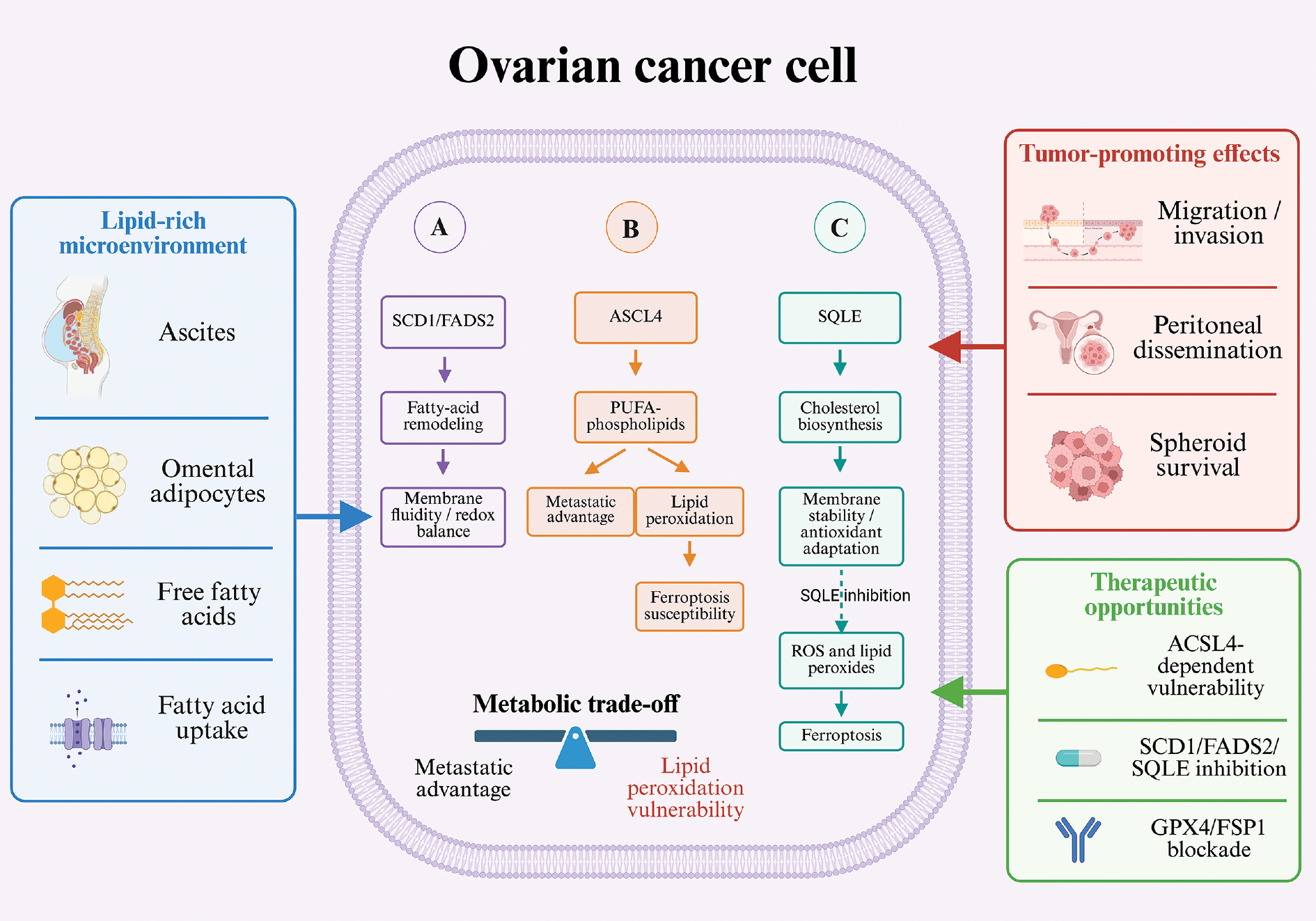
The lipid composition in cell membranes establishes the substrate basis for LP. Acyl-CoA synthetase long-chain family member 4 (ACSL4) promotes the incorporation of PUFAs, including arachidonic and adrenic acids, into membrane phospholipids and is a major determinant of ferroptosis susceptibility. At the same time, ACSL4 and polyunsaturated lipids can increase membrane fluidity and facilitate tumor-cell deformation, extravasation, and metastatic colonization [6]. This creates a clinically relevant metabolic trade-off: ACSL4 activity and PUFA-phospholipid enrichment may indicate both enhanced metastatic fitness and a greater potential oxidative burden. Whether this state becomes therapeutically exploitable depends on the capacity of the tumor to continuously detoxify lipid peroxides.
Fatty acid desaturation by stearoyl-CoA desaturase 1 (SCD1) and fatty acid desaturase 2 (FADS2), together with changes in cholesterol metabolism, can further influence membrane properties and redox balance. These metabolic features are best interpreted alongside direct measurements of lipid composition and oxidation products rather than as standalone markers of ferroptosis susceptibility.
For patient stratification, lipidomics should assess not only total fatty-acid abundance but also PUFA-containing phospholipids, the balance of mono- and polyunsaturated lipids, oxidized phospholipids, and cholesterol-related membrane features. Because primary tumors, ascites-derived cells, and omental metastases may differ in lipid exposure and membrane oxidizability, biomarker validation should include paired sites whenever feasible (Figure 1).
Figure 1 Lipid metabolic adaptation and ferroptosis vulnerability in ovarian cancer. The lipid-rich peritoneal microenvironment promotes fatty acid uptake and membrane remodeling, which supports tumor-cell survival and peritoneal dissemination while increasing the abundance of oxidizable phospholipids. The resulting ferroptotic vulnerability is determined by the balance between lipid substrate availability and antioxidant defense pathways, including GPX4- and FSP1-mediated protection. Figure created in BioRender (Yang, X. 2026; https://BioRender.com/bm4tbk9).
Activation of ferroptosis defense pathways is not equivalent to functional dependence
The SLC7A11–GSH–GPX4 axis is the principal system for removing phospholipid hydroperoxides. SLC7A11 sustains cystine uptake and GSH synthesis, whereas GPX4 uses GSH to reduce phospholipid hydroperoxides to nontoxic lipid alcohols. In ovarian cancer, CCAAT/enhancer-binding protein gamma (CEBPG) has been shown to upregulate SLC7A11 transcription [7]. This example illustrates how ferroptosis defenses can be regulated, but expression changes alone do not establish whether a tumor depends on the pathway for survival.
FSP1–CoQ10 and NRF2-regulated programs provide parallel or compensatory defenses. A noncanonical FSP1-dependent ferroptosis escape pathway has also been implicated in platinum resistance in ovarian cancer [8]. Such findings highlight the possibility that FSP1- and NRF2-associated defenses can compensate for impaired GPX4 activity. Consequently, the more informative marker is not the abundance of an individual defense protein, but evidence that the tumor relies on the broader defense network for survival.
Biomarker assessment must distinguish pathway expression from activity and, crucially, from functional dependence. Molecular profiling can characterize expression, while GSH status, CoQ10 redox balance, NRF2 target activity, and oxidized phospholipids may indicate pathway activity. Dependence, however, requires evidence that targeted inhibition causes LP-associated cell death in patient-derived models and that ferroptosis-suppressing treatment reverses this effect.
A biomarker-guided framework for patient stratification
On the basis of current evidence, we propose a three-dimensional framework that requires prospective validation: the burden of peroxidizable lipid substrates, the level of ongoing LP pressure, and the functional reserve of ferroptosis defense systems. These dimensions are not interchangeable. High PUFA-phospholipid abundance does not by itself establish ferroptotic sensitivity. Increases in 4-HNE or malondialdehyde (MDA) may represent nonspecific oxidative injury. Likewise, high GPX4 or FSP1 expression may indicate pathway activation without proving dependence. The most plausible therapeutic window exists when a high substrate burden, active LP stress, and experimentally verifiable defense dependence converge (Table 1 and Figure 2).
Table 1 Core Stratification Axes, Contextual Modifiers, and Safety Gate for LP-Guided Patient Stratification in Ovarian Cancer
| Framework Role/Assessment Dimension | Candidate Indicators | Suggested Samples/Technologies | Clinical Interpretation and Limitations |
|---|---|---|---|
| Core axis 1: peroxidizable lipid substrate | PUFA phospholipids, ACSL4-associated lipid features, membrane unsaturation | Lipidomics of tissue or ascites-derived tumor cells; mass spectrometry imaging | Indicates abundant oxidizable substrates but does not independently establish treatment sensitivity |
| Core axis 2: existing LP pressure | Oxidized phospholipids, 4-HNE, C11-BODIPY signal, labile iron | Fresh samples, tissue staining, flow cytometry, or imaging | Requires multiple convergent markers; MDA or total ROS should remain supportive evidence only |
| Contextual marker: defense pathway status | SLC7A11, GPX4, FSP1, NRF2, and downstream targets | Immunohistochemistry, transcriptomics, proteomics, and metabolic assays | Reflects expression or activity but cannot be equated directly with functional dependency |
| Core axis 3: functional defense dependence | LP and cell death after GPX4, FSP1, or another candidate defense-pathway inhibitor, with quantified rescue by mechanistically distinct ferroptosis-suppressing interventions | Patient-derived tumor cells, ascites-derived spheroids, organoids, or fresh tissue slices | Core treatment gate; most closely linked to predicted response, but provisional thresholds require prospective calibration |
| Contextual modifier: spatial and temporal heterogeneity | Differences in lipid composition, LP pressure, and defense dependence among primary tumors, ascites-derived tumor cells, metastatic lesions, and recurrent lesions, including changes before and after treatment | Paired sampling, longitudinal biopsy, single-cell profiling, spatial profiling, and repeated ascites sampling | Quantifies cross-site and longitudinal discordance; does not by itself redefine patient status |
| Safety gate: safety and immune context | Sensitivity of normal tissues and immune cells to pathway inhibition | Immunocompetent models, peripheral blood, and tumor-immune profiling | Separate safety gate that defines whether a biologically eligible tumor can be treated within an acceptable therapeutic window |
Figure 2 Operational framework for LP-guided patient stratification in ovarian cancer. Paired samples inform three core axes: the lipid substrate burden, LP pressure, and functional defense dependence, with functional dependence serving as the treatment gate. A prespecified index lesion or compartment defines patient-level status, and other paired sites are used to quantify spatial heterogeneity. Figure created in BioRender (Yang, X. 2026; https://BioRender.com/y5904w5).
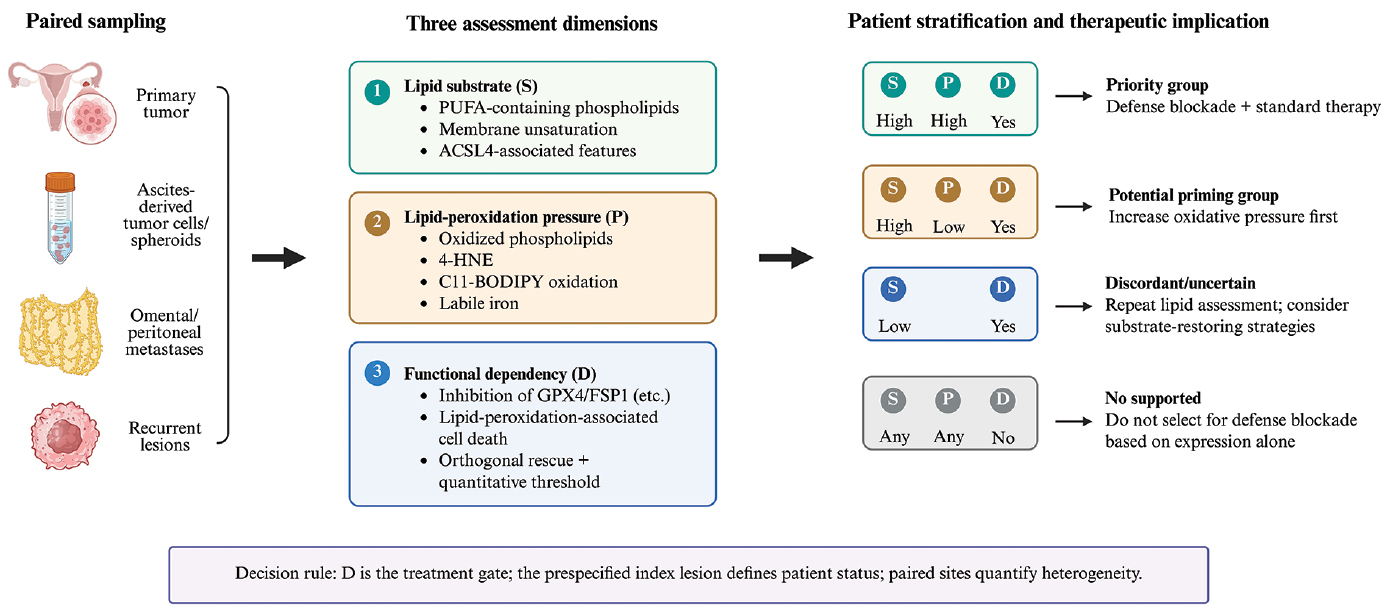
For prospective pilot studies, the framework needs to be sufficiently operational to test validated clinical score while remaining clearly distinct from it. Assay-specific, cohort-based thresholds may therefore be more appropriate than fixed clinical cutoffs at this stage. A high substrate burden could initially be defined by PUFA-phospholipid abundance in the upper quartile of the study cohort, while high LP pressure could require concordant elevation of at least two indicators, such as oxidized phospholipids, 4-HNE, C11-BODIPY oxidation, or labile iron. Functional dependence would act as the treatment gate and should be supported by quantitative inhibition-and-rescue experiments using at least two mechanistically distinct ferroptosis-suppressing interventions. For pilot work, a prespecified rescue fraction, such as a reduction of at least 50% in treatment-induced excess cell death, could serve as a provisional research threshold and then be recalibrated prospectively. A minimal panel could thus combine targeted lipidomics, one tissue-based or cellular marker of LP, and one quantitative functional assay. The same prospective logic should be used when several lesions are available from the same patient. An index lesion or compartment should be designated before biomarker testing, preferably a recurrent or progressing lesion sampled close to the treatment decision; when tissue is unavailable, contemporaneous malignant ascites may serve as an alternative index compartment. Other paired sites can then be used to measure concordance and heterogeneity rather than to redefine patient status retrospectively.
Application of these rules yields four provisional patient categories. Tumors with a high substrate burden, high LP pressure, and confirmed functional dependence represent the most straightforward candidates for direct disruption of anti-LP defenses in combination with standard treatment. A second group has a high substrate burden and confirmed dependence but relatively low current pressure, which suggests that oxidative priming with platinum agents, poly(ADP-ribose) polymerase (PARP) inhibitors, or lipid remodeling strategies may be needed before defense pathways are blocked. A third, discordant group has a low substrate burden despite confirmed dependence. Rather than being combined with nondependent disease, this pattern should prompt repeat or orthogonal lipid assessment and, in research settings, evaluation of whether substrate-restoring strategies alter sensitivity. The remaining group lacks experimentally confirmed dependence regardless of substrate or pressure status and should not be selected for ferroptosis defense blockade solely because a pathway marker is highly expressed. In every category, biological eligibility remains subject to a separate safety gate.
This classification is a translational research framework rather than a clinical scoring system. Future composite models may integrate lipidomics, spatial localization, protein expression, and functional assays, but they must be validated in independent cohorts and demonstrate predictive value beyond single-marker testing for GPX4, SLC7A11, or FSP1. Cross-site discordance should therefore be reported explicitly as part of the biological phenotype rather than compressed retrospectively into a single patient label.
Clinical settings that should be prioritized for stratification studies
Patients with malignant ascites and extensive peritoneal dissemination
Tumor cells in ascites are directly exposed to a lipid-rich, low-adhesion, and nutritionally fluctuating environment, so they are an informative material for evaluation of metabolic adaptation and ferroptosis defense dependence. Ascites can often be sampled repeatedly and can support lipidomic analysis, short-term cultures, spheroid assays, and organoid-based functional drug testing. However, as the ascitic environment itself can modify ferroptotic sensitivity, the way in which these samples are handled becomes part of the assay. Removal of tumor cells from ascites may change both LP pressure and defense dependence. Whenever feasible, intact spheroids should therefore be tested in autologous ascites or matched ascites-conditioned conditions alongside parallel cultures after ascites removal. Comparison of these conditions can show whether ascites primarily suppresses LP pressure, alters GPX4/FSP1 dependence, or affects both. If ascites-derived cells differ from the primary tumor, that result should be interpreted as complementary information about disseminated disease rather than as a reason to over-ride the prespecified index lesion automatically.
Patients with platinum- or PARP inhibitor–resistant disease
Treatment-resistant disease may provide a useful setting in which to test this framework, but resistance itself should not be taken as evidence of ferroptosis sensitivity. Platinum agents increase oxidative stress in addition to causing DNA damage, and some resistant cells may survive by strengthening detoxification of lipid peroxides, which can create a potentially targetable dependence. It has been reported that shikonin enhances cisplatin activity together with ferroptotic features [9], but its broad, multitarget pharmacology means that this effect cannot be attributed specifically to ferroptosis [10]. PARP inhibitors may also modify ferroptotic pressure: niraparib restricts intraperitoneal metastasis through CD36-dependent ferroptosis, while sphingosine kinase 1 (SPHK1) enhances olaparib resistance through the nuclear factor kappa B (NF-κB)–NRF2–ferroptosis pathway [11, 12]. Taken together, these observations support platinum- or PARP inhibitor–resistant disease as a setting for biomarker enrichment and hypothesis testing, while functional confirmation remains necessary before this approach can be used for treatment selection.
Histologic subtypes with distinct metabolic backgrounds
Epithelial ovarian cancer subtypes may differ fundamentally in lipid metabolism, iron homeostasis, and antioxidant circuitry. Ovarian clear-cell carcinoma has a distinctive redox-adapted phenotype, and ferroptosis has been implicated in eribulin-induced cytotoxicity in this subtype [13]. Histology may therefore provide a rational starting point for enrichment, but it cannot replace functional testing. Even within the same subtype, dependence may vary between patients and among lesions within the same patient.
From biomarkers to clinical trials: how should the stratification strategy be validated?
Before a stratification strategy can be tested clinically, ferroptosis needs to be measured in a consistent way. Total reactive oxygen species and MDA are useful indicators of oxidative injury, but neither is specific enough to establish ferroptotic cell death. Preclinical studies should combine lipid-selective oxidation assays with measurements of oxidized phospholipids or 4-HNE, labile iron, relevant defense proteins, and compatible morphologic changes. The same principle applies to functional rescue. Rescue by ferrostatin-1 alone is not mechanistically definitive because radical-trapping antioxidants act at the LP level and can suppress other forms of peroxidative injury [14]. Stronger evidence would come from reproducible rescue via mechanistically distinct ferroptosis-suppressing interventions, such as a radical-trapping antioxidant together with an iron-chelating intervention, combined with parallel assessment of apoptosis and necrosis. These orthogonal controls are particularly important when patient-derived material is used, for which general oxidative toxicity could otherwise be mistaken for ferroptosis.
Turnaround time is just as important as analytical depth. In recurrent disease, a result that takes several weeks may arrive too late to guide treatment. A workable clinical pathway could begin with a focused panel based on lipidomics, immunohistochemistry, or both. For tumors that appear to carry a high LP burden, short-term testing could then be performed in fresh explants, primary cultures, or organoids. The interpretation of these assays also needs to reflect the current therapeutic landscape. Clinically validated, selective GPX4- or FSP1-directed therapeutics are not yet available, and dedicated ferroptosis-directed agents remain at an early stage of clinical translation [15]. Functional testing therefore currently provides evidence of biological dependence rather than an immediately actionable drug-selection result. A staged approach of this type is less comprehensive than unrestricted multiomics but is more compatible with the time available for treatment decisions.
These practical constraints also shape how the biomarker strategy should be tested clinically. Biomarker-enriched studies are useful at an early stage because they can establish assay feasibility, pharmacodynamic target engagement, and preliminary antitumor activity, but alone they cannot show that the biomarker predicts treatment benefit. Once feasibility is established and a clinically suitable ferroptosis-directed agent becomes available, the next step should be a biomarker-stratified randomized study that includes both biomarker-positive and biomarker-negative patients. Within each stratum, standard therapy alone could be compared to standard therapy plus the ferroptosis-directed intervention for direct testing of biomarker-by-treatment interactions. Paired pretreatment and on-treatment tumor or ascites samples could then be used to assess oxidized phospholipids, 4-HNE, and defense pathway activity to help in distinguishing inadequate drug exposure from failure of the stratification model itself.
The therapeutic window may represent the major limitation of ferroptosis-targeted therapy. GPX4 is not a tumor-specific survival factor but an essential safeguard against membrane LP in normal tissues. Genetic inactivation of Gpx4 in mice causes acute renal failure, and renal sensitivity to Gpx4 loss may differ according to sex and NRF2-mediated antioxidant capacity [16, 17]. GPX4 is also required for maintenance of follicular helper T cells, so systemic GPX4 inhibition might compromise immune-cell survival and function [18]. Moreover, GPX4-associated ferroptosis has been implicated in cisplatin-induced cochlear injury, which highlights the auditory system as another potential site of vulnerability to excessive ferroptotic stress [19]. Systemic GPX4 inhibition could therefore result in organ toxicity and weakening of immune-mediated tumor control.
The safety consequences of targeting FSP1 and NRF2 are less clearly defined and are likely to be tissue- and context-dependent. Nevertheless, both pathways contribute to cellular protection against oxidative injury, and simultaneous disruption of several ferroptosis defense systems may further narrow the therapeutic window. Clinical development should therefore demonstrate a meaningful difference in dependence between tumor and normal tissues rather than assume that greater oxidative stress in cancer cells is sufficient for selectivity. Preclinical studies should compare the exposure required for antitumor activity with the exposure that causes renal, hepatic, hematologic, auditory, and immune toxicity, and assess whether treatment impairs T-cell function. Local delivery, intermittent dosing, or targeting of tumor-enriched upstream regulators may reduce systemic exposure, but these approaches require direct experimental validation and should not be regarded as substitutes for establishing tumor selectivity.
Conclusion
The value of LP for precision treatment of ovarian cancer lies not in the possibility of another universal cell-death therapy, but in identification of a subset of tumors with a definable metabolic and defense dependence. A lipid-rich peritoneal environment can support metastatic fitness while at the same time shaping latent ferroptotic vulnerability; whether that vulnerability becomes clinically useful depends on the relationship among the peroxidizable lipid burden, ongoing LP pressure, and functional defense dependence in the disease compartment most relevant to treatment. Future work should therefore combine paired sampling, lipidomics, and quantitative functional assays while preserving the biological context of ascites and explicitly reporting cross-site discordance. Clinical validation can then progress from biomarker-enriched feasibility studies to biomarker-stratified randomized trials that formally test biomarker-by-treatment interactions. In this way, patient stratification would become part of the development path for ferroptosis-based therapy rather than an additional step considered only after such therapies reach the clinic.
Data availability statement
Data and materials can be provided on reasonable request to one of the corresponding authors.
Ethics statement
No direct interactions with human or animal subjects were involved. Therefore, ethical approval and informed consent were not required.
Author contributions
Shaojie Zhao and Yan Zhang conceptualized the review. Xinfeng Yang, Yida Wang, and Haiyue You wrote the manuscript. Xinfeng Yang prepared the figures and the table. Shaojie Zhao and Yan Zhang critically reviewed and edited the manuscript. Yan Zhang and Shaojie Zhao obtained funding support. All authors read and approved the final manuscript.
Acknowledgments
The authors acknowledge BioRender.com for the graphical resources used to create the illustrations in this manuscript.
Funding
The work was supported by the National Natural Science Foundation of China (grant no. 82472842), the Medical Key Strategic Project Fund of Wuxi Health Commission, and the Wuxi “Taihu Light” Science and Technology Program (Medical and Health Technology Project, grant no. Y20242116).
Conflict of interest
The authors declare that there are no conflicts of interest.
Consent for publication
Not applicable.
References
- Chen F, Kang R, Tang D, Liu J. Ferroptosis: principles and significance in health and disease. J Hematol Oncol 2024;17(1):41. [PMID: 38844964 DOI: 10.1186/s13045-024-01564-3]
- Kapper C, Oppelt P, Arbeithuber B, Gyunesh AA, Vilusic I, et al. Targeting ferroptosis in ovarian cancer: novel strategies to overcome chemotherapy resistance. Life Sci 2024;349:122720. [PMID: 38762066 DOI: 10.1016/j.lfs.2024.122720]
- Liu D, Hu Z, Lu J, Yi C. Redox-regulated iron metabolism and ferroptosis in ovarian cancer: molecular insights and therapeutic opportunities. Antioxidants (Basel) 2024;13(7):791. [PMID: 39061859 DOI: 10.3390/antiox13070791]
- Ruan D, Wen J, Fang F, Lei Y, Zhao Z, et al. Ferroptosis in epithelial ovarian cancer: a burgeoning target with extraordinary therapeutic potential. Cell Death Discov 2023;9(1):434. [PMID: 38040696 DOI: 10.1038/s41420-023-01721-6]
- Setayeshpour Y, Chen SY, Dayanidhi DL, Lee Y, Wu SC, et al. Ascites protects against ferroptosis and enables the peritoneal growth of ovarian cancer. Nat Commun 2026;17(1):4190. [PMID: 42115216 DOI: 10.1038/s41467-026-72116-1]
- Wang Y, Hu M, Cao J, Wang F, Han JR, et al. ACSL4 and polyunsaturated lipids support metastatic extravasation and colonization. Cell 2025;188(2):412-29.e27. [PMID: 39591965 DOI: 10.1016/j.cell.2024.10.047]
- Zhang X, Zheng X, Ying X, Xie W, Yin Y, et al. CEBPG suppresses ferroptosis through transcriptional control of SLC7A11 in ovarian cancer. J Transl Med 2023;21(1):334. [PMID: 37210575 DOI: 10.1186/s12967-023-04136-0]
- Zhao Z, Ge G, Gong M, Liu Y, Ma L, et al. Redox-triggered USP18 confers cisplatin resistance in ovarian cancer by selectively activating a non-canonical FSP1-dependent ferroptosis escape pathway. Redox Biol 2026;93:104179. [PMID: 42030646 DOI: 10.1016/j.redox.2026.104179]
- Ni M, Zhou J, Zhu Z, Xu Q, Yin Z, et al. Shikonin and cisplatin synergistically overcome cisplatin resistance of ovarian cancer by inducing ferroptosis via upregulation of HMOX1 to promote Fe2+ accumulation. Phytomedicine 2023;112:154701. [PMID: 36773431 DOI: 10.1016/j.phymed.2023.154701]
- Lew CY, Tang YT, Lee AY, Chin ZJ, Chang WL, et al. Shikonin as a dietary phytochemical with multi-target anti-cancer activities: from molecular mechanisms to translational applications. Nutrients 2025;17(19):3085. [PMID: 41097161 DOI: 10.3390/nu17193085]
- Jin N, Qian YY, Jiao XF, Wang Z, Li X, et al. Niraparib restricts intraperitoneal metastases of ovarian cancer by eliciting CD36-dependent ferroptosis. Redox Biol 2025;80:103528. [PMID: 39922130 DOI: 10.1016/j.redox.2025.103528]
- Teng K, Ma H, Gai P, Zhao X, Qi G. SPHK1 enhances olaparib resistance in ovarian cancer through the NFκB/NRF2/ferroptosis pathway. Cell Death Discov 2025;11(1):29. [PMID: 39875359 DOI: 10.1038/s41420-025-02309-y]
- Azumi M, Kusama K, Yoshie M, Nakano S, Tsuru A, et al. Involvement of ferroptosis in eribulin-induced cytotoxicity in ovarian clear cell carcinoma. Eur J Pharmacol 2024;971:176544. [PMID: 38552939 DOI: 10.1016/j.ejphar.2024.176544]
- Zilka O, Shah R, Li B, Friedmann Angeli JP, Griesser M, et al. On the mechanism of cytoprotection by ferrostatin-1 and liproxstatin-1 and the role of lipid peroxidation in ferroptotic cell death. ACS Cent Sci 2017;3(3):232-43. [PMID: 28386601 DOI: 10.1021/acscentsci.7b00028]
- Zayed M, Massoud A, Elwakeel E, Ezzat P, Alammar Y, et al. Repurposing approved drugs as ferroptosis modulators: a critical review of clinical trials in cancer and neurodegeneration. Naunyn Schmiedebergs Arch Pharmacol 2026;399(12):18655-72. [PMID: 42319414 DOI: 10.1007/s00210-026-05582-5]
- Friedmann Angeli JP, Schneider M, Proneth B, Tyurina YY, Tyurin VA, et al. Inactivation of the ferroptosis regulator Gpx4 triggers acute renal failure in mice. Nat Cell Biol 2014;16(12):1180-91. [PMID: 25402683 DOI: 10.1038/ncb3064]
- Ide S, Ide K, Abe K, Kobayashi Y, Kitai H, et al. Sex differences in resilience to ferroptosis underlie sexual dimorphism in kidney injury and repair. Cell Rep 2022;41(6):111610. [PMID: 36351395 DOI: 10.1016/j.celrep.2022.111610]
- Yao Y, Chen Z, Zhang H, Chen C, Zeng M, et al. Selenium-GPX4 axis protects follicular helper T cells from ferroptosis. Nat Immunol 2021;22(9):1127-39. [PMID: 34413521 DOI: 10.1038/s41590-021-00996-0]
- Liu Z, Zhang H, Hong G, Bi X, Hu J, et al. Inhibition of Gpx4-mediated ferroptosis alleviates cisplatin-induced hearing loss in C57BL/6 mice. Mol Ther 2024;32(5):1387-406. [PMID: 38414247 DOI: 10.1016/j.ymthe.2024.02.029]