Smart Hydrogels Empowering Tissue Repair: Material Design, Emerging Applications, Repair Mechanisms, and Future Challenges
1State Key Laboratory of Frigid Zone Cardiovascular Diseases (SKLFZCD), Department of Pharmacology, College of Pharmacy, Harbin Medical University, Heilongjiang 150081, PR China
2Department of Inorganic Chemistry and Physical Chemistry, College of Pharmacy, Harbin Medical University, Heilongjiang 150081, PR China
3Research Unit of Health Sciences and Technology (HST), Faculty of Medicine, University of Oulu, Finland
4Department of Pharmaceutics, College of Pharmacy, Harbin Medical University, Daqing Campus, Heilongjiang 163319, PR China
5Key Laboratory of Basic Research and Health Management on Chronic Diseases in Heilongjiang Province, Harbin Medical University, Heilongjiang 163319, PR China
*Correspondence to: Yang Li, E-mail: liy@hrbmu.edu.cn, Yang.Li@oulu.fi; Na An, E-mail: 1164958668@qq.com; Ting Zhang, E-mail: tingzhang@hrbmu.edu.cn
Received: January 5 2026; Revised: March 17 2026; Accepted: April 12 2026; Published Online: May 15 2026
Cite this paper:
Gong Y, Sun B, Xiao X et al. Smart Hydrogels Empowering Tissue Repair: Material Design, Emerging Applications, Repair Mechanisms, and Future Challenges. BIO Integration 2026; 7: 1–26.
DOI: 10.15212/bioi-2026-0004. Available at: https://bio-integration.org/
Download citation
© 2026 The Authors. This is an open access article distributed under the terms of the Creative Commons Attribution License (https://creativecommons.org/licenses/by/4.0/). See https://bio-integration.org/copyright-and-permissions/
Abstract
Smart hydrogels are attracting considerable interest in the biomedical field, because of their high water content, excellent biocompatibility and biodegradability, and distinctive properties in response to stimuli. Their three-dimensional mesh structure effectively mimics the microenvironment of human tissue, by maintaining moist conditions conducive to wound healing while also serving as a support for active ingredients, thus ensuring their precise and controlled release. Consequently, these materials have excellent potential for use in tissue regeneration. This article first classifies smart hydrogels according to their response mechanisms, specifically systems that respond to temperature, pH, light, and magnetic fields. It then systematically examines potential applications of smart hydrogels in tissue regeneration, according to their ability to dynamically adapt to different tissue microenvironments, particularly the regeneration of skin, bone and cartilage, nerve tissue, and internal organs. Despite their promising potential, smart hydrogels still face several challenges, including imbalances in the rates of tissue degradation and regeneration, insufficient mechanical properties, and relatively limited functionality. Future research should focus on material modification and optimization, AI-assisted design, and interdisciplinary collaboration between medicine and engineering to develop hydrogels’ multifunctional, personalized integration and clinical application, and ultimately enable smarter, more effective solutions for tissue engineering and regenerative medicine.
Keywords
Drug delivery, mechanism of action, smart hydrogels, stimuli responsive, tissue regeneration.
Introduction
Tissue damage and dysfunction pose serious health problems. Wound healing is a complex and organized pathophysiological process comprising several phases, which are generally classified into thrombosis; inflammation; new tissue formation (including re-epithelialization and granulation tissue formation); and tissue remodeling and resorption [1–3]. Because severe tissue damage substantially affects patients’ physical and mental well-being, appropriate therapeutic approaches must be developed [4]. Numerous biomaterials have been demonstrated to accelerate tissue repair with greater efficacy than drugs in certain clinical applications, thus prompting substantial interest [5]. Hydrogels are among these materials. Their characteristic three-dimensional structure allows hydrogels to store large amounts of water [6]. In addition, hydrogels are hydrophilic, flexible, and biocompatible, and they have adaptable mechanical properties, controlled degradation profiles, and high reactivity. They can mimic the state of human tissue, maintain a moist environment, and serve as drug carriers and thus promote wound healing [7–10].
The glucan-based hydrogel developed by Shen et al., with quaternary ammonium functional groups and adjustable cross-linking density, has been shown in in vivo animal models to significantly shorten wound healing time [11]. Because its excellent water retention properties help maintain a moist environment that effectively promotes wound healing, this material is suitable for modern dressings. Simultaneously, the porous structure of the hydrogel, its swelling ability, and its reactivity to stimuli provide an effective system for drug delivery. Modified hydrogels provide delayed drug release, thus enabling decreased frequency of administration and improved patient compliance. In addition, modification of the hydrogel surface facilitates targeted delivery [12, 13]. He et al. have reported microscopic analysis indicating that quaternary chitosan (QCS)/oxidized branched polysaccharide hydrogels, with different polymer ratios, have the same porous structure, which favors the storage of drugs, nutrients, and metabolic by-products [14]. The sericin- and flax-based nanocomposite hydrogel reported by Rafat et al. is a drug delivery system with many hydroxyl functional groups that can absorb drugs [15]. It maintains therapeutic concentrations in wounds through self-diffusion and therefore has application potential in wound dressings. Because of their excellent fluidity, adhesiveness, and degradability, certain hydrogels can adapt to irregularly shaped wounds and adhere firmly to the surrounding tissue [16, 17] before being gradually absorbed and degraded by the body. Arguchinskaya et al. have implanted a hydrogel structure synthesized from high-substitution gelatin methacrylate (GelMA) under the skin in mice and observed its structural degradation over time [18]. The hydrogel adhered firmly to the surrounding tissue and degraded slowly without causing a notable inflammatory reaction.
Hydrogels, by combining exceptional physicochemical properties with biological functionality, have considerable potential in the tissue engineering and regeneration fields [8]. The main advantages of hydrogels are as follows. First, their high water content creates a local moist environment mimicking the conditions of human tissue and creating conditions conducive to wound healing [19]. Moreover, their excellent biocompatibility decreases the risk of immune rejection and effectively resolves immune compatibility issues in clinical tissue reconstruction through tissue engineering [20]. Their adaptation flexibility allows for specific microenvironments to be created as needed, which can be tailored to the requirements of different reconstruction scenarios [21]. Their degradability allows them to be absorbed by the body without requiring additional surgical intervention to remove them, thereby greatly improving patient adherence to treatment [18, 22]. Beyond offering these numerous advantages, smart hydrogels also exhibit specific and controllable responses to stimuli and respond to various external influences (e.g., temperature, pH, and light), and therefore can adapt to different environments [23, 24]. These fundamental advantages serve as the basis for the wide application of hydrogels, particularly smart hydrogels, in tissue regeneration. This review focuses on the classification of smart hydrogels; their applications and advanced mechanisms in various fields of tissue regeneration; current issues and obstacles; and future prospects.
Material design of smart hydrogels
Hydrogel classification and crosslinking methods
Hydrogels can be classified according to their origin, cross-linking method, and reactivity to external stimuli (Figure 1). Hydrogels can be divided into natural hydrogels and synthetic hydrogels according to their origin. Natural hydrogels consist of natural biomacromolecules and are a rich source of materials with excellent biocompatibility and biodegradability [25]. These materials primarily include polysaccharides such as sodium alginate, hyaluronic acid (HA), cellulose, and chitosan, as well as protein substances such as gelatin and collagen [25, 26]. Chitosan-based hydrogels have a wide range of applications in wound treatment because of their excellent biocompatibility, hemostatic properties, oxygen permeability, and antimicrobial effects [27]. Methyl acrylate gelatin hydrogels retain the biocompatibility of natural gelatin while further enhancing material crosslinking stability, and therefore provide potential tissue engineering materials suitable for 3D cell culture and 3D bioprinting [28]. Synthetic hydrogels are prepared from polymeric materials; common synthetic precursors include polyethylene glycol (PEG), polyvinyl alcohol (PVA), and polyacrylamide [29]. In related application studies, Dobner et al. first demonstrated that a non-degradable PEG-based hydrogel, delivered via injection to ischemic myocardium, delays post-infarction left ventricular dilatation [30]. Moreover, Gong et al.’s phytic acid polyvinyl alcohol hydrogel, using a synergistic hydrogel delivery plus drug therapy approach, has achieved superior repair outcomes in corneal injury models [31]. Natural hydrogels and synthetic hydrogels, beyond having distinct origins, substantially differ in several key properties. From the viewpoint of biocompatibility, natural hydrogels are obtained from natural biomolecules (e.g., polysaccharides or proteins), and their chemical structure is highly similar to that of the extracellular matrix (ECM). They promote cell adhesion, migration, and proliferation; are easily recognized and accepted by human cells; and generally exhibit excellent biocompatibility [32, 33]. However, because synthetic hydrogels do not possess biological activity of their own, their biocompatibility has been questioned [34]. Whereas natural hydrogels, in contrast, can be broken down via physiological enzymatic processes in the body [32, 33], synthetic hydrogels are not biodegradable on their own, and degradable bonds must be used to control their rate of decomposition [33]. From the viewpoint of chemical resistance and mechanical properties, natural hydrogels have low chemical resistance and poor mechanical properties and stability [35]. In contrast, synthetic hydrogels are precisely engineered through advanced polymer chemistry, and therefore have high chemical resistance and exceptional mechanical properties [34, 35]. These two types of hydrogels considerably differ in key characteristics, such as their origin, composition, biocompatibility, and degradability. The choice or modification of compounds should be based on the specific requirements of the biomedical application.
Figure 1 Schematic diagram of core attributes of smart hydrogels. This diagram illustrates key characteristics of smart hydrogels across three core dimensions: source, crosslinking method, and stimulus responsiveness. Source is categorized as natural (collagen, cellulose, proteins) or synthetic (polyethylene glycol (PEG), polyvinyl alcohol (PVA), and polyacrylamide (PAM)). Crosslinking method encompasses physical crosslinking (intermolecular forces without chemical bond formation) and chemical crosslinking (reaction-mediated). Stimulus responsiveness covers response types such as magnetic fields, light, pH, temperature, and related applications (e.g., drug delivery), and corresponding benefits are noted for selected response types. Created in BioRender. S, B. (2025) https://BioRender.com/dgyzmei.
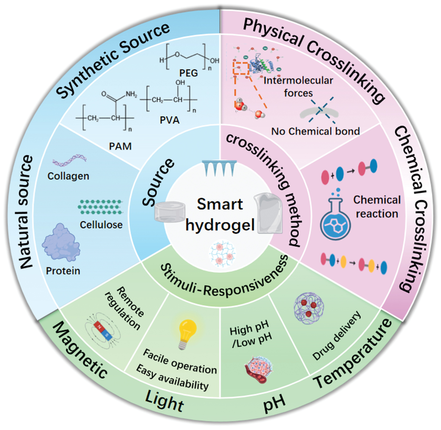
Hydrogels can be categorized by cross-linking method into physically cross-linked hydrogels and chemically cross-linked hydrogels, according to whether covalent bonds are formed. Physically crosslinked hydrogels are formed via intermolecular forces without chemical reactions, and the resultant physical crosslinking is intrinsically reversible [25]. These intermolecular interactions include hydrogen bonding, electrostatic forces, and hydrophobic interactions [36]. The non-covalent interactions within polyelectrolyte complexes are a key mechanism for hydrogel formation. For example, Potaś et al. have demonstrated that chitosan (CS, positively charged) and sodium alginate (negatively charged) spontaneously form a polyelectrolyte complex suitable for hydrogel-based dressings, via electrostatic interactions between the amino group at the C2 position of CS’s glucosamine unit and the carboxyl group of sodium alginate [37]. Chemically crosslinked hydrogels form covalent bonds through reactions such as click chemistry [38], enzymatic reactions [39], radical polymerization [40], and Schiff base reactions [41]. Compared with physically cross-linked hydrogels, chemically cross-linked hydrogels have a more stable structure, excellent mechanical properties, and controllable degradation characteristics [6, 25]. Li et al. have summarized click chemistry applications in biomedical hydrogels and highlighted that hydrogels produced through this strategy are widely used in key areas such as drug delivery, cell culture, tissue regeneration, biosensor fabrication, and 3D bioprinting [42].
In hydrogel production, the choice of materials is critical. With advances in modern science and technology, designing hydrogel materials via artificial intelligence (AI) has become an innovative approach to design and selection, and is increasingly used in hydrogel development. This method primarily involves a combination of machine learning (ML) and 3D bioprinting to predict, filter, and refine the three-dimensional models needed for experimental prototype design [43]. Compared with the previous traditional prediction–design–optimization–experiment method, this method has high precision and efficiency, and it enables cost savings. Concrete models proposed on the basis of 3D bioprinting combined with ML can improve hydrogel design approaches. Sokmen et al. have proposed combining AI and 3D bioprinting technology to design structures for coronary arteries [44]. ML, a fundamental technology in the field of AI, includes various disciplines such as control theory, deterministic theory, mathematical statistics, and computer science. It is also integrated into various application areas such as hydrogel design and breast cancer screening [45, 46]. ML solves complex problems associated with the selection of the appropriate types and number of building blocks, such as monomers, in the design of hydrogel-based materials, as well as the diversity of these components in the material. By generating many design models by using permutations and combinations and then performing algorithmic analysis to exclude irrelevant models, this approach decreases reliance on traditional trial-and-error experiments and enables more precise and effective hydrogel design. For example, in fields such as drug delivery and the development of biological ink, AI has enabled revolutionary advances in the hydrogel design and optimization through ML [43, 47]. Liao et al. have used a combined approach including ML, intelligent data analysis, and experimental research to examine a database of more than 20,000 amino acid sequences of adhesive proteins [48]. The identified characteristic sequences suitable for underwater adhesives have facilitated the development of super-sticky hydrogels for wet environments. On the basis of these sequence characteristics, the authors have predicted and developed approximately 180 types of hydrogels and created a reference database for the ML-based intelligent design–prediction–optimization process. This framework served as the basis for the development of ML algorithms for the development of hydrogels with excellent adhesive properties under conditions of high humidity. AI-based methods for material selection can therefore contribute to the development of smart hydrogels.
Stimulus-responsive hydrogels
Reactive hydrogels are considered smart biomaterials characterized by dynamic interactions and promising applications in tissue regeneration. These hydrogels dynamically react to various stimuli, such as temperature, pH, ions, and light, and subsequently undergo changes in their physical and chemical properties, particularly mechanical strength, biocompatibility, volume, and the release of active substances [49].
Temperature-responsive hydrogels
Because of their typical thermosensitive properties, thermosensitive hydrogels are highly promising in wound treatment and drug delivery. Cai et al. have developed a multifunctional reactive hydrogel based on carboxymethylagarose and n-isopropylacrylamide, whose thermosensitive properties generate a contraction force at 30°C that improves wound healing [50]. In addition, owing to the interactions among the polymers, and between the polymers and water, this hydrogel enables controlled delivery at different temperatures. Zhang et al. have presented a thermosensitive injectable hydrogel system filled with hybrid levobupivacaine and poly(D,L-lactide)-poly(ethylene glycol)-poly(D,L-lactide) (hLB/PLEL) that remains liquid at room temperature [51]. After administration in the body, it transforms into a semi-solid hydrogel under the influence of physiological temperature fluctuations, thus ensuring prolonged release of levobupivacaine and significantly prolonging the local anesthetic effect. The organic hydrogel developed by Gui and colleagues combines thermosensitive mechanical properties with a programmable shape memory function that is temperature dependent [52]. This hydrogel not only has excellent thermotherapeutic effects but also releases anti-inflammatory drugs and cools inflamed wounds; therefore, it provides an ideal thermosensitive anti-inflammatory dressing. These hydrogels have shown potential in synergistic therapies and clinical applications, because of their ability to accurately detect thermal signals, thus providing excellent opportunities for development and research in smart biomedical materials.
Light-responsive hydrogels
Light is an intuitively understandable signal that can be manipulated to regulate various stimulating signals in time and space. Intelligent control of hydrogels by using light has become an important field of research. Wang et al. have synthesized a protein-based photosensitive hydrogel that depends on vitamin B12 and, under light exposure, rapidly changes from a gelatinous state to a solution state, while its polymeric component, C-terminal adenosylcobalamin binding domain (CarHc), rapidly decomposes [53]. This light-sensitive gel system promotes accelerated release/restoration of stem cells and protein molecules. Wang et al. have developed and synthesized a light-sensitive HA hydrogel for the dynamic immunomodulation of macrophages [54]. This new strategy for controlling inflammatory responses accelerates regeneration of endogenous tissue and therefore has considerable potential for tissue regeneration. Fan et al. have developed pyramid-shaped microneedle systems (photoresponsive drug delivery microspheres-integrated pyramid microneedle systems, abbreviated as PDDM-MN) equipped with light-sensitive microspheres containing drugs [55]. They have excellent photothermal properties and reproducible reactivity in the near-infrared range. Animal studies have demonstrated the ability of these innovative and superior drug delivery systems to achieve controlled insulin release and regulate blood glucose levels in mice with streptozotocin-induced diabetes.
pH-responsive hydrogels
pH-responsive hydrogels react to microenvironmental characteristics such as the low pH of diabetic wounds, thereby enabling controlled drug release, gel degradation, and microenvironmental increases [56]. The pH/magnetic dual-responsive hemicellulose-based nanocomposite hydrogel developed by Long et al. has demonstrated excellent controlled release properties for both acetylsalicylic acid and theophylline, and therefore might serve as a potential carrier for targeted drug delivery, particularly under gastrointestinal conditions [57]. Park et al. have developed a pH-responsive hydrogel based on carboxymethyl cellulose/hydroxyethyl acrylate (CMC/HEA), featuring a stable network structure and favorable mechanical properties [58]. The new cl-CMC-g-pHEA hydrogel acts as a transdermal delivery system for naringenin, which can be used to treat various skin lesions caused by pH imbalance. Wu et al. have developed an injectable polylysine-based glucopeptide hydrogel that reacts to pH and reactive oxygen species and has excellent antioxidant and antibacterial properties [59]. Under conditions of low pH and high activity of reactive oxygen species, which are characteristic of diabetes, this hydrogel exhibits dual reactivity and promotes the spatiotemporal release of the angiogenesis stimulator mangiferin and the anti-inflammatory compound diclofenac sodium. This innovation provides a promising dressing material for accelerating the healing of chronic wounds caused by diabetes. Yi and colleagues have presented a new two-layer hydrogel, with basic fibroblast growth factor (bFGF) and 4-hydroxyphenylboronic acid pinacol ester (PAPE)-modified fucoidan/chitosan/morin nanoparticles (CFMNPs) [60]. Because a specific pH-dependent degradation mechanism regulates drug release, this hydrogel can adapt to the dynamic physiological environment during various phases of ulcer healing and has been demonstrated to be highly effective in the clinical treatment of burns and frostbite.
Magnetically responsive hydrogels
Magnetically sensitive hydrogels enable regulation of release processes by using remotely controlled magnetic fields. Hia and colleagues have developed a material called magnetically controlled hydrogel (McSa@m-Sp), a composite in which sodium alginate serves as a matrix to incorporate superparamagnetic iron oxide nanoparticles coated with calcium phosphate [61]. This material offers a controlled degradation rate, strong antibacterial effects, osteogenic properties, and selective release of active ingredients, and therefore has promise in bone engineering. Xue and colleagues have integrated apoptosis-modified PEG/polyethylene imide and apoptosis-modified palladium-hydrogen-based nanocatalysts into a polyacrylamide/HA double-network hydrogel [62]. This hydrogel exhibits excellent sensitivity to wireless magnetic fields and promotes intervertebral disc regeneration by stimulating cell absorption, proliferation, and differentiation. Manescu Paltanea et al. have demonstrated that magnetic hydrogels have promising potential in complex multimodal tumor therapies and enhanced immunotherapies [63]. These applications are useful because of their distinctive functional advantages, such as controlled magnetic injection, synergistic shear stress reduction properties, and magnetically tuned and enhanced mechanical properties.
Cutting-edge applications and mechanisms of hydrogels in diverse tissue engineering repair scenarios
Skin tissue repair
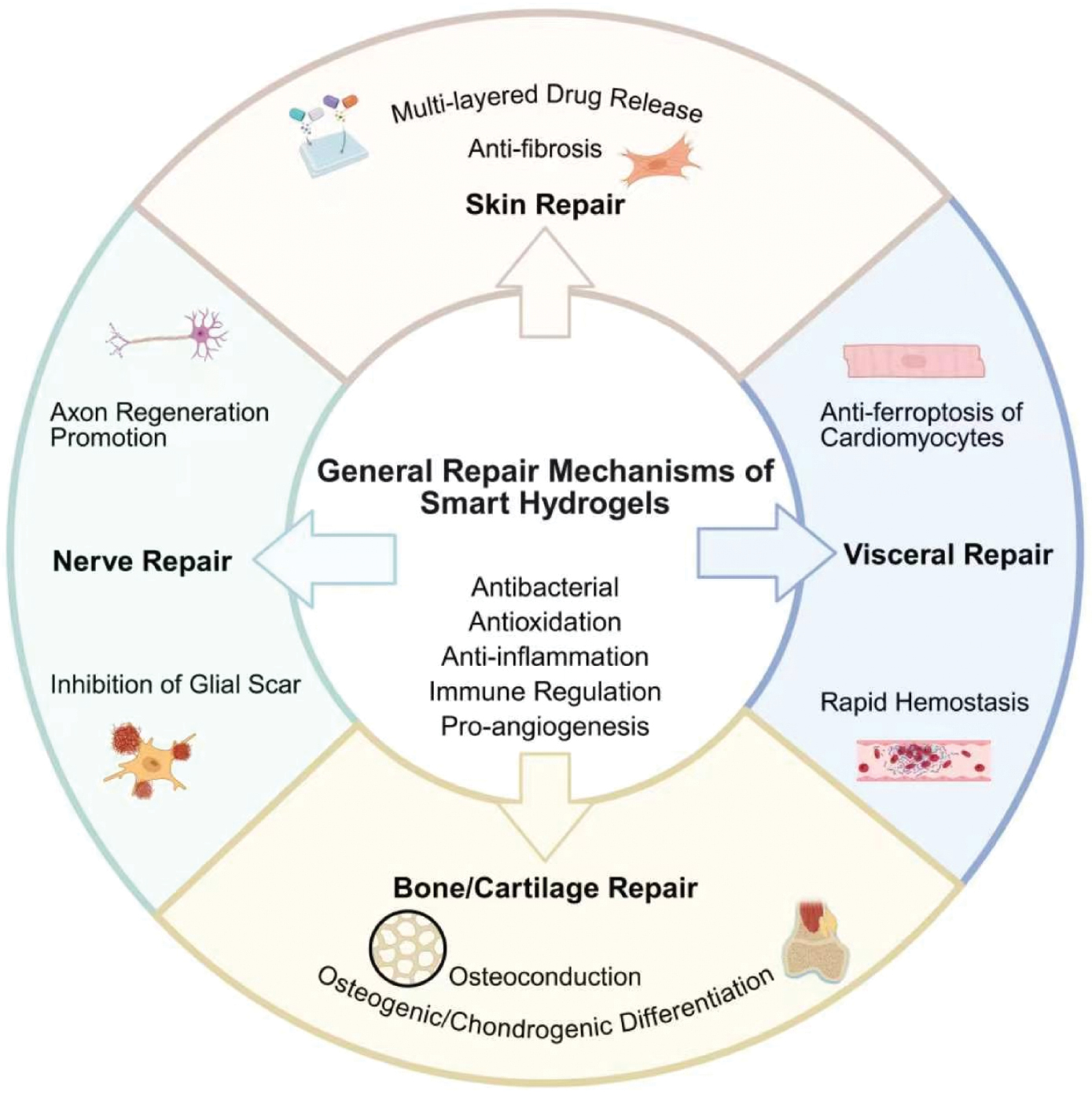
The general repair mechanisms of smart hydrogels and their applications in various tissue repair scenarios are summarized in Figure 2. Hydrogels offer numerous intriguing possibilities for promoting skin healing. Examples include antimicrobial, conductive, and self-healing hydrogel dressings [64]; micro-needle hydrogel dressings filled with microbubbles containing mitochondria-rich stem cells to promote chronic wound healing [65]; and cross-linked polysaccharide-based bilayer hydrogels that accelerate skin healing [66]. Although these materials share common advantages, their properties also differ. Most feature high antimicrobial activity, excellent conductivity, superior self-healing properties, strong adhesion to tissue, favorable biocompatibility, high hemostatic effects, potent antioxidant activity, and strong anti-inflammatory effects. The two-layer crosslinked structure further enhances mechanical properties while ensuring partial breathability at the wound site. Other hydrogels achieve high degradability and absorbency through the addition of supplementary substances. However, most hydrogels also have limitations. Attempts to incorporate substances such as polyamide PA to improve specific properties often introduce compromises that weaken other aspects and hinder optimal functionality. Furthermore, some processes require high-precision equipment. Existing technical limitations, such as the production of magnetic nanoparticles for hydrogels, hinder increases and prevent the optimization of test results. Additionally, highly complex manufacturing processes increase costs and therefore hinder clinical application. Hydrogels offer diverse applications in skin tissue regeneration. Researchers are leveraging the numerous advantages of hydrogels to effectively treat wounds such as burns, chronic wounds (diabetic wounds), and acute trauma. Current advanced applications focus on developing innovative hydrogels incorporating other substances, thereby providing diverse and enhanced functions.
Figure 2 Schematic illustration of the general repair mechanisms of smart hydrogels and their applications in tissue regeneration. Smart hydrogels exert therapeutic effects via core universal mechanisms (antibacterial, antioxidative, anti-inflammatory, immunoregulatory, and pro-angiogenic activities), and are applied in four key tissue repair scenarios: skin repair (multi-layered drug release and anti-fibrosis), visceral repair (rapid hemostasis and anti-ferroptosis of cardiomyocytes), bone/cartilage repair (osteogenic/chondrogenic differentiation, and osteoconduction), and nerve repair (axon regeneration promotion and glial scar inhibition). Created in BioRender. Siqi, S. (2026) https://BioRender.com/81u1066.
Application scenarios
Burn injuries
Burns are common injuries in everyday life, usually caused by high temperatures, electric shock, radiation, or chemicals [67]. Deep burns are prone to infection and disrupted cell function, and can elicit excessive inflammatory responses [68]. Because they lack the components necessary for local regeneration, these extensive skin injuries often result in excessive hypertrophic scars or severe fibrosis, which compromises aesthetic outcomes [69]. Delayed healing due to immunosuppression and metabolic reactions in patients increases the risk of complications such as sepsis [70]. To remedy these problems, Gong et al. have developed a hygroscopic antibacterial hydrogel containing a network of curcumin and polyphenol-magnesium (Cur-Mg@PP) [71]. By leveraging interactions within the hydrogel to improve the stability of curcumin and the properties of curcumin, Mg2+, and PP, the authors developed a highly adaptable moisturizing hydrogel capable of releasing bioactive substances in a well-protected form over a long time period. This hydrogel effectively adapts to the shape of burns and promotes healing.
Chronic wounds
Chronic wounds (such as diabetic ulcers) are difficult to treat and, in severe cases, can lead to amputation or death. As society advances, the demand for effective treatments for chronic wounds is growing. However, excessive immune responses in people with diabetes considerably impede wound healing. Existing pharmacological treatments are not only limited in their effectiveness and expensive but also are suitable only for small wounds [72]. Therefore, research on treatments for chronic wounds continues. Zhang et al. have developed a vascular hydrogel (F/R gel) capable of undergoing proportional and dynamic cross-linking reactions [73]. In the treatment of chronic wounds, such as those associated with diabetes, vascular diseases, and pressure ulcers, healing is difficult and often stalls in the infection or severe inflammation phases. This hydrogel’s multilayer release properties, excellent adhesion, and self-repair capabilities regulate healing in a programmed manner and adapt to the irregular surface of the wound. Its injectability, self-repair capability, and adhesion enable adaptation to the size and surface of the wound by adjustment of the injection volume. In addition, the F/R gel’s adaptive function enables modulation of immune and fibroblastic subtypes, thereby promoting antimicrobial control of infections, angiogenesis, adequate ECM deposition, and suppression of skin fibrosis. Its inhibitory effect on Gram-negative and Gram-positive bacteria, together with its biocompatibility, contributes to rapid epithelialization and suppression of chronic wound healing.
Acute injury
Acute injuries are often accompanied by spinal cord injury. Existing treatment methods, including surgical resection, require administration of high doses of glucocorticoids to suppress inflammation but simultaneously trigger immune reactions in other tissues. Patients who receive intensive treatment with corticosteroids and antibiotics after surgery experience considerable physical stress. These interventions delay healing and can sometimes be harmful to the body [74]. To address this problem, Wang et al. have developed polylysine-based hybrid hydrogels (PBHEVs@AGN) [75]. These hydrogels consist of internal aminoguanidine (AGN) nanoparticles, which are rapidly released in response to pH changes, and extracellular vesicles, which are slowly released over time. Blockade of the TLR4/Myd88/NF-κB inflammatory pathway creates a stable anti-inflammatory microenvironment for the synergistic treatment of acute and subacute spinal cord injuries (SCIs). Simultaneously, the PBHEVs@AGN delivery system shows lasting restoration of motor function, high tissue preservation, diminished scarring, and increased regeneration of myelin and axons, all of which favor SCI treatment and scar-free restoration.
Scar-free repair
Many advanced-stage wounds heal poorly and form hypertrophic scars. Infections associated with burns can lead to excessive scarring and fibrosis, whereas chronic inflammatory reactions can lead to scarring due to sensory nerve damage, excessive collagen deposition, and excessive fibroblast proliferation. Traditional dressings, such as gauze or membrane materials, serve primarily as barriers preventing the entry of new bacteria. However, they contribute little to tissue regeneration or scar prevention [76, 77]. Existing clinical methods for decreasing scarring, such as laser therapy or cryotherapy, provide some improvements, yet their effectiveness is limited. Therefore, new treatment methods are continually being developed [78]. Yang et al. have proposed mesenchymal stem cells (MSCs), specifically a hydrogel containing human umbilical cord mesenchymal stem cell (HucMSC) exosomes, as well as a hydrogel filled with HucMSCs [79]. In this hydrogel, DP7/Exo is crucial in manifesting anti-inflammatory effects by regulating macrophage polarization and the expression of inflammatory cytokines. HucMSC-Exo DP7/Exo decreases total wound healing time and suppresses collagen deposition, thereby allowing wounds to heal without scarring. Specifically, exosomes (HucMSC-Exos) were encapsulated in the macroporous HA hydrogel HD-DP7/Exo and lyophilized for long-term storage. When used, they rapidly released miR-21–5p into healing cells, dissolved, and subsequently exerted therapeutic effects. Zhao and colleagues have developed a bioactive hydrogel adhesive dressing with excellent adherence to moist tissue [80]. The rapid gelation process is enabled by an internal prepolymer, poly(quinic acid)-PEG-g-dopamine, and amino-terminated Pluronic F127 (APF) micelles loaded with astragaloside IVwith amino termination. APF micelles significantly decrease the swelling coefficient of the hydrogel, and internally generated H2O2 residues have strong antimicrobial effects. This hydrogel with added micelles exhibits injectability, high strength, and anti-edema properties, and achieves rapid hemostatic effects. It is also highly effective for closing skin incisions without sutures and for healing infected skin wounds without leaving scars.
Mechanisms of action
Hydrogels have many applications in skin tissue repair and are suitable for various types of wounds, including difficult-to-heal pathological wounds. For example, they greatly accelerate healing of wounds typical of diseases such as diabetes. Their exceptional ability to regenerate skin arises from their excellent biocompatibility, antimicrobial activity, antioxidant properties, regulation of inflammation, and stimulation of angiogenesis. These characteristics allow hydrogels to effectively modulate the expression of various cytokines, reactive oxygen species (ROS), and other substances in the body, thereby unleashing their powerful regenerative abilities.
Antibacterial properties
Park et al. have investigated hydrogels containing tannic acid (TA) within double-crosslinked hydrogels composed of PVA [81]. TA induces bacterial enzyme complexation through its astringent properties after binding various biomolecules. Concurrently, it inhibits oxidative phosphorylation in bacterial membranes, blocks bacterial bioactivity, and consequently exerts antibacterial effects. Sun et al. have proposed incorporating borax into a PVA/polyacrylic acid (PAA) hydrogel network via swelling and partial dehydration [82]. Free boron ions released during hydrogel swelling disrupt bacterial cell membranes and induce quorum sensing effects, thus achieving nearly 99.99% killing rates against Escherichia coli and Staphylococcus aureus through this ion-release mechanism.
Antioxidant properties
Guo et al. have reported that the injectable hydrogel QCS/TA, which contains QCS and TA, has active groups that directly bind free radicals, according to fuchsin and DPPH experiments [83]. Its excellent free radical scavenging ability determines its superior antioxidant ability. The glucose-sensitive hydrogel platform HA methacrylate-PBA/catechin developed by Xu et al. was based on HA and reacted with a molar equivalent amount of DPPH solution (200 mM) in methanol [84]. Components such as catechin within the hydrogel scavenge free radicals and decrease ROS levels.
Regulating inflammatory responses
Song et al. have developed an injectable thermosensitive hydrogel composed of ECM hydrogel@exosomes (ECM@exo) derived from cardiomyocytes [85]. Immunohistochemical staining and qRT-PCR analysis indicated that ECM@exo decreases interferon tumor necrosis factor-α (TNF-α) and interleukin (IL-6) expression, thereby mitigating inflammatory responses. Concurrently, miRNAs within exosomes modulate immune cell differentiation and development, activate inflammatory signaling pathways, and regulate inflammatory responses. Liu et al. have developed a hybrid hydrogel assembled from bis-synthesized glycopeptides [86]. Immunofluorescence staining experiments observing macrophage infiltration have demonstrated that this hydrogel polarizes primary macrophages recruited to wound sites into the M2 type. Furthermore, it promotes the secretion of anti-inflammatory cytokines (IL-10) and transforming growth factor (TGF), thereby repairing the inflammatory microenvironment of the wound.
Promoting angiogenesis
Jin et al. have developed a patch based on a GelMA-diacetylated starch (DAS)-citrus exosome hydrogel (GelMA/DAS/Exo-hydrogel) by incorporating citrus exosomes into a hydrogel composed of GelMA and DAS [87]. Tubular structure formation experiments and WB analysis of Arg-1 and iNOS protein expression revealed that components within citric acid exosomes considerably elevated expression of the vascular genes CD31 and vascular endothelial growth factor A (VEGFA), thus demonstrating the GelMA/DAS/Exo hydrogel’s role in promoting vascular repair. A polypyridine-HA (Ppy-HA) hydrogel formulated by Chu et al. has been found to promote angiogenesis by inducing M2 macrophage polarization and subsequent release of cytokines including VEGF, TGF-β, and platelet-derived growth factor [88].
Bone and cartilage tissue repair
Hydrogels also have multiple innovative applications in stimulating bone and cartilage regeneration. The most notable examples include biomimetic mineralized hydrogels for bone defect repair [89], thermosensitive hydrogels filled with MSCs for cartilage defect repair [90], and dual-network hydrogels that improve the mechanical support of cartilage regeneration [91]. These applications have many advantages. For example, they are highly adaptable, because they can adjust to the mechanical and biochemical microenvironment of bone and cartilage, while allowing relatively free regulation. They are highly functional, because they can absorb both cells and factors, thus achieving dual structure and regulatory functions. They are highly biocompatible: most hydrogel materials (e.g., HA or chitosan) have low immunogenicity and therefore avoid rejection reactions. However, they also pose certain problems. For example, their mechanical properties are limited; even with the support of a double network, hydrogels used for cartilage tissue regeneration are often insufficiently strong and cannot withstand repeated friction in joint cavities. The timing between hydrogel degradation and tissue regeneration remains suboptimal; a mismatch between the rate of degradation and rate of new bone tissue formation can impair regeneration. Some hydrogels have low toxicity because of the growth factors that they contain, thereby decreasing the effectiveness of regeneration and increasing the burden on the body. In addition, some latest-generation hydrogels pose clinical application problems. For example, their complex and costly large-scale production might prevent their widespread introduction into clinical practice. Precisely because of the aforementioned advantages, hydrogels are currently used to repair various bone and cartilage lesions in various situations. Current research is aimed at improving the mechanical properties of hydrogels and achieving high tissue compatibility by adding additional materials to form stable structures, thereby contributing to the development of various innovative hydrogels for clinical applications.
Application scenarios
Irregular bone defects
Bone irregularities generally vary in shape, size, and depth, and are common in patients with diabetes. Although autogenous bone grafts remain the standard treatment for bone reconstruction, their clinical use poses serious problems because of their limited availability and high morbidity at donor sites. Most current clinical treatment strategies focus on improving one or more capabilities but have disadvantages in other areas (e.g., adhesion to irregular surfaces). In dentistry, irregular cranial and facial bone defects affect patient aesthetics, which is a complex but urgent problem [92, 93]. Zhou et al. have developed an injectable, wireless, ultrasound-activatable, bone-adhering nanocomposite hydrogel [93]. In this hydrogel, amine-modified piezoelectric nanoparticles form dynamic covalent bonds with the hydrogel’s biopolymer network. These dynamic bonds confer injectability, adaptability, and electromechanical responsiveness. In addition, amino-modified barium titanate nanoparticles (KBTO) improve the adhesion strength between KBGO and irregular bone surfaces. Under the influence of ultrasound, hydrogel promotes osteoblast differentiation and accelerates bone healing in vitro; this process is mediated by the ERK/MAPK and PI3K-AKT signaling pathways in response to electrical stimulation.
Diabetic bone defects
Diabetic bone abnormalities are bone lesions characterized by elevated volume, diminished quality, susceptibility to infection, and chronic inflammation. Altered macrophage metabolism prevents the release of inflammatory factors and the maintenance of proinflammatory states; simultaneously, elevated AFK levels and altered glucose metabolism modify bone metabolism. Decreased stem cell activity in affected areas prevents osteoblast differentiation and hinders bone defect repair [94, 95]. Zhang et al. have developed a factor-independent reactive hydrogel that combines ROS-destructible thionic bridges (TK) and UV-responsive norbornene (NB) groups with eight-arm PEG macromolecules, thus resulting in UV-induced gelation [95]. The hydrogel rapidly transforms into a gel under UV exposure and then decomposes. Over time, it promotes the migration and infiltration of bone marrow MSCs (BMSCs) while removing ROS, thereby facilitating the transition of BMSCs from the adipogenic differentiation pathway to the osteogenic differentiation pathway. The main advantage of this hydrogel is its degradation mechanism, which eliminates ROS and therefore the need to introduce exogenous growth factors. This approach solves the problems of growth factor toxicity and additionally improves bone healing in patients with diabetes.
Osteoporotic bone defects
Osteoporosis is a systemic bone disease characterized by a markedly loss of bone mass leading to bone fragility and changes in bone microarchitecture. Osteoporotic bone defects in patients with osteoporosis are often caused by trauma. For example, postmenopausal osteoporosis is among the most common skeletal diseases in women. Treatment of these defects is aimed at stimulating bone angiogenesis. However, modern clinical treatment methods often involve substances such as growth factors that cause adverse effects [96, 97]. To solve this problem, Wei has developed a biomimetic hydrogel [97]. The S-nitrozoglycine and Ca2+ contained in this hydrogel stimulate the release of biologically active nitric oxide by BMSCs and human vascular endothelial cells, and activate the NO/cGMP signaling pathway, thereby improving communication between osteogenesis and angiogenesis. The hydrogel improves stem cell homeostasis by including the SKPPGTSS peptide and modulates the microenvironment for bone defect healing by regulating matrix stiffness, thus conferring immunomodulatory properties. To improve biocompatibility and targeting, the sodium alginate (SA) was modified with dopamine and chelated calcium ions to increase stiffness.
Osteoarthritis
Osteoarthritis is a disease of the cartilage tissue characterized by premature wear of the cartilage surface and often accompanied by degeneration of the articular cartilage and abnormal remodeling of the subchondral bone. Restoring the metabolic homeostasis of cartilage and bone and effective penetration into the subchondral bone are currently considered important therapeutic targets in the treatment of osteoarthritis [98, 99]. Yang and colleagues have developed a cross-linked hydrogel with multiple hydrogen bonds filled with TA and chondrogenic factor (KGN) [100]. This structure confers stable mechanical properties and ensures gradual release of the active ingredient. The hydrogel can withstand 28,000 loading and unloading cycles, and it has rapid shape memory at body temperature. Because the developed network remains stable under constant mechanical stimulation, it might have future use prospects in minimally invasive surgery. In addition, the sequential release of TA and KGN promotes the differentiation of MSCs from bone marrow into chondrocytes. PTK hydrogels incorporating both KGN and TA within the PMI hydrogel network demonstrate high tissue adhesion. In addition, they mitigate local inflammatory responses and eliminate ROS, thereby creating an optimal microenvironment for the migration of BMSCs and promoting cartilage regeneration throughout its entire thickness.
Osteosarcoma
Osteosarcoma, the most common primary malignant bone cancer, affects primarily children and adolescents and causes movement disorders. This cancer is caused by the uncontrolled proliferation of tumor cells and osteolysis in the affected area. Clinical treatment therefore focuses on postoperative anatomical reconstruction and the prevention of recurrence in the affected area. Strategies for postoperative rehabilitation in osteosarcoma are currently being researched to develop approaches that restrict tumor growth and repair bone defects [101, 102]. Chu and colleagues have developed a bioactive nanocomposite hydrogel that prolongs the release of bioactive magnesium ions, antibodies to programmed death-1 (PD-L1), and vismodegib [103]. This approach eliminates remaining tumor cells while modulating the immune responses of sentinel lymph nodes and consequently improving immune reactivity. Simultaneously, the released magnesium ions promote the osteogenic differentiation of BMSCs and further support bone regeneration in defects after osteosarcoma resection.
Mechanism of action
Hydrogels importantly can promote bone tissue regeneration. Because of their excellent biocompatibility and mechanical properties, they can firmly bind bone tissue and subsequently facilitate the restoration of irregular bone defects. In addition, hydrogels’ biological activities promote bone formation, stimulate vascular regeneration, and have anti-inflammatory effects and immunomodulatory properties. Consequently, these materials can stimulate or suppress the production of certain substances in the body, thereby facilitating the repair of severe bone and cartilage defects that are difficult to treat.
Osteogenic bioactivity
The dynamic DNA/GelMA hydrogel (CGDE) developed by Zhu et al. is characterized by high expression of osteogenic proteins [104]. The CGDE matrix improves intracellular expression of the transcription factors RUNX2 and OSX while stimulating the secretion of type I collagen (COL-1) and osteocalcin, thus conferring high biocompatibility and osteogenic bioactivity. Wang et al. have developed a Ca2+-TA-rGO (CTAG/GelMA) hydrogel incorporating nanolayers of oxidized graphene (GO) and TA (TA-rGO), which introduce Ca2+ into GelMA [105]. This hydrogel ensures sustained release of Ca2+ and promotes osteogenic differentiation of MSCs. It plays a regulatory role in the expression of several key osteogenic-related factors, including osteomorphogenin-2 (BMP-2), osteocalcin, and osteopontin. Simultaneously, it elevates the concentration of extracellular calcium ions (Ca2+), thereby further promoting the uptake and utilization of calcium ions by cells or tissues. This hydrogel also considerably enhances the expression of ALP in BMSCs from culture with M2 macrophages and promotes osteogenic differentiation. In addition, it further improves stability by inhibiting osteoclast differentiation.
Promoting vascular regeneration
Bai and others have conducted experiments indicating that demineralized bone matrix and Zn2+, present in the hydrogel of their multifunctional biomimetic structure, stimulate the secretion of angiogenic factors, such as VEGF [106]. Deferoxamine (DFO) activates the proangiogenic HIF-1 signaling pathway, thereby improving vascular regeneration. Human urinary stem cell-derived exosomes (USCEXOs) have been developed by Lu et al. USCEXOs/G5H2, formed by the conjugation of HA methacrylate and nano-hydroxyapatite, stimulates angiogenesis through the high expression of CD31, HIF1A, and fibroblast growth factor (FGF1) [107].
Anti-inflammatory effects
The curcumin-quaternized chitosan/pluronic F (Cur-QCS/PF) hydrogel developed by Chen et al. releases curcumin while inhibiting the expression of inflammatory cytokines such as IL-1β and TNF-α [108]. Consequently, inhibition of inflammation and ECM destruction in tendon tissue results in anti-inflammatory effects. A GelMA/Met@ZIF-8 hydrogel (imidazolium zeolite structure filled with metformin) developed by Lao et al. releases both metformin and zinc ions [109]. This hydrogel has anti-inflammatory effects by regulating macrophage polarization through immunomodulation, decreasing the M1 phenotype and inflammatory mediators (inducible nitric oxide synthase, iNOS, IL-1), and increasing the M2 phenotype markers (arginase-1, Arg-1) and inflammatory mediators (TNF-β). Simultaneously, the contained hypoglycemic drug metformin has anti-inflammatory effects by decreasing inflammatory cytokine expression, thus contributing to positive therapeutic outcomes.
Immunomodulatory effects
Luo et al. have developed the CSBDX@MOF system by using Mg-GA MOF with immunomodulatory activity [110]. Under the action of 4-formylphenylboronic acid (4-FPBA), dynamic boron-imine bonds form with dextran (DEX) and carboxymethylchitosan (CMCS), in an interpenetrating network structure (CMCS/4-FPBA/DEX, CSBDX) and a CSBDX@MOF system containing magnesium and gallic acid (GA). Mg-GA MOF has immunomodulatory effects. The components Mg2+ and GA decrease the production of proinflammatory mediators and increase the M2/M1 ratio. The DEX contained in the hydrogel acts synergistically with Mg2+ and GA by inhibiting the M1-mediated increase in IL-6, COX-2, and iNOS while promoting secretion of TGF-β3 and IL-10 by M2 macrophages, thereby attenuating inflammation. Wei et al. have synthesized mesoporous silica nanoparticles (MSNs) filled with s-nitroglycerin (s-NG) by encapsulating cell membranes obtained from MSCs of bone marrow and human umbilical vein endothelial cells [97]. MSNs filled with GSNO were synthesized through the encapsulation of cell membranes derived from BMSCs and human umbilical vein endothelial cells on the MSN surface, thus yielding SA-MSN@CM-Stiff. Flow cytometry analysis and western blotting experiments indicated that this formulation suppresses the formation of cluster of differentiation 86 positive (CD86+) macrophages and simultaneously promotes the formation of CD206+ macrophages. Polarized macrophages markedly increase expression of the angiogenic factor VEGF-A and the bone formation-related molecule BMP-2, thereby exerting immunomodulatory effects by regulating macrophage polarization.
Neural tissue repair
Hydrogels are highly biocompatible and biomimetic materials that can mimic the microscopic environment of tissue during nerve regeneration and encourage nerve cell adhesion, migration, and growth. In addition, hydrogels can enable targeted delivery of bioactive substances and provide the signaling molecules necessary for nerve regeneration. Therefore, hydrogels are often used in the treatment of injuries or diseases that affect the central or peripheral nervous system, and their importance as an adjunct to traditional surgical procedures is increasing.
Application scenarios
Repair of central nervous system injuries
Damage to the central nervous system can be categorized into two main types: traumatic brain injury and traumatic SCI [111]. The main challenge in treating these injuries is the limited regenerative ability of mature neurons in the brain; consequently, injuries often lead to permanent functional disability. To address this fundamental problem, Li et al. have proposed a treatment strategy using hydrogels that react with and trap ROS [112]. This strategy is aimed at facilitating spinal cord repair. Taking advantage of the hydrogel’s excellent biocompatibility and its specific responsiveness to ROS, the researchers successfully and precisely delivered BMSCs in vitro to the site of SCI. This selective degradation of excess ROS decreased oxidative damage at the injury site, decreased the expression of inflammatory mediators such as IL-1β/IL-6/TNF-α, and suppressed glial and fibrotic scar formation, thus creating a microenvironment favoring neural recovery. Simultaneously, the hydrogel protected the encapsulated BMSCs and decreased cell death at the injury site. In this way, BMSCs can promote interneuron formation and regenerate axons through paracrine effects: the secretion of neurotrophic factors (such as bFGF and glial cell-derived neurotrophic factor) and the modulation of macrophage polarization toward anti-inflammatory M2 cells. Consequently, recovery outcomes and motor function restoration improve after SCI. For recovery after traumatic brain injury, Zhang et al. have proposed a hydrogel composite (BGA@GelMA) [113]. By mimicking the micro- and nanostructure as well as the mechanical properties of the brain’s ECM, it forms a biomimetic three-dimensional structure that provides attachment points for human neural progenitor cells (hNPCs). It then continuously releases brain-derived neurotrophic factor (BDNF), glial cell-derived neurotrophic factor, and cyclic adenosine monophosphate at the site of traumatic brain injury, thus meeting the need for acute repair. Simultaneously, it activates the mitogen-activated protein kinase (MAPK) signaling pathway and stimulates glutamate secretion, thereby significantly improving the expression of neuronal markers such as TUJ1 and MAP2 in hNPCs. hNPCs in BGA@GelMA hydrogels, compared with Matrigel, show higher gene expression associated with neuronal differentiation. Consequently, the induction of targeted differentiation of hNPCs into functional cortical intermediate neurons restores the damaged area of the brain. In addition, hydrogels can serve as nerve electrodes for brain-computer interfaces. Khan et al. have proposed hydrogel nerve electrodes capable of continuously monitoring brain nerve signals for 8 weeks [114]. In experiments on rats that had experienced strokes, these electrodes regulated damaged neurons, decreased areas of cerebral infarction, and promoted the restoration of motor function, thus offering new possibilities for restoring damaged nerve connections and monitoring nerve information in the brain.
Interventions for central nervous system degenerative diseases
Degenerative diseases affecting the central nervous system are a group of disorders caused by nerve cell degeneration and leading to a decline in cognitive, motor, and other functions [115]. The development of these diseases is usually associated with genetic factors, environmental influences, and abnormal protein accumulation. For example, the pathological features of Alzheimer’s disease include β-amyloid accumulation around nerve fibers and neurofibrillary tangles caused by tau protein hyperphosphorylation [116]. The prevalence of Alzheimer’s disease increases with age. After diagnosis, patients gradually lose the ability to care for themselves in the final stage of the disease and experience severely impaired quality of life in older age. Because no cure currently exists for this disease, early detection and treatment remain key strategies for slowing its progression. Liu et al. have developed a thermosensitive hydrogel (BP-MB@Gel) containing phosphorus and methylene blue nanoparticles [117]. This hydrogel is injected intranasally, and its heat-sensitive properties cause it to quickly change from a liquid to a gel at the optimal temperature of the nasal mucosa. Consequently, BP-MB remains in the nasal cavity and is continuously released for 48 hours. When BP and MB reach the brain, they synergistically eliminate ROS, inhibit tau protein phosphorylation, and protect mitochondria. This method decreases neurofibrillary tangles and simultaneously increases ATP production, thereby improving cognitive function. Parkinson’s disease (PD), a neurodegenerative disorder that affects primarily older people, is characterized by the progressive degeneration and death of dopaminergic neurons in the substantia nigra, which in turn significantly decrease dopamine levels in the brain [118]. Motor disorders as well as non-motor symptoms, such decreased sense of smell and autonomic dysfunction, may result. Because the disease progresses slowly, continuously, and irreversibly, early diagnosis and intervention are crucial for alleviating PD symptoms and slowing its progression. Chen et al. have coated polyurethane nanoparticles with polydopamine (PUD) and combined them with chitosan and ethylene glycol to create a biodegradable and flexible hydrogel (CPUD-gel) [119]. After implantation, this hydrogel mimics the properties of brain tissue, has anti-inflammatory and antioxidant effects, promotes neuron proliferation, supports neuronal differentiation, and accelerates the regeneration of dopaminergic neurons. Research in mice with PD has indicated that CPUD gel, when combined with acupuncture therapy, significantly improves treatment outcomes and motor performance.
Repair of peripheral nervous system injuries
Damage to the peripheral nervous system is associated with damage to the brain and spinal cord caused by trauma, pressure, metabolic disorders, and other factors. These injuries are usually classified as traumatic and non-traumatic [120]. The main difference with respect to central nervous system damage is the ability of nerve cells to regenerate close to the damaged area. However, regenerated axons that lack appropriate guidance signals tend to deviate from their intended path, thus affecting functional recovery. To solve this problem, Xu et al. have proposed an electrically controlled hydrogel channel made of highly sensitive nanofibers [121]. In the body, it generates electrical stimulation induced by external ultrasound waves, which selectively stretches the nerve cells and stimulates axon growth. Simultaneously, the heat-sensitive hydrogel in the outer layer shrinks and releases nerve growth factors into the intercellular space, thus further accelerating peripheral nerve regeneration. Lee et al. [122] have developed a new neuroinductive catheter (PGC) whose outer layer uses poly(L-lactide-co-lactide) to mimic the nerve epithelium. The inner gelatin hydrogel is cross-linked with visible light during 3D bioprinting to create surface patterns with microstriae and Arg-Gly-Asp sequences, which improve nerve cell adhesion. It also absorbs nerve growth factor and ensures its gradual release for as many as 58 days through swelling and enzymatic degradation. Its effectiveness in promoting axon regeneration and restoring motor function has been demonstrated in a rat model with sciatic nerve injury.
Mechanism of action
Hydrogels meet a wide range of requirements for restoring damaged nerves in various conditions and have achieved highly effective results in post-traumatic treatment. They replicate the microenvironment within the nerve and precisely control it to promote recovery. Their main advantage is their ability to mimic the microenvironment of nerves and precisely regulate it for regeneration. Hydrogels can decrease inflammatory reactions, prevent glial scar formation, and create a favorable environment for nerve regeneration. In addition, hydrogels can provide the structural support necessary for nerve cell regeneration and use several mechanisms to support effective regeneration.
Resisting inflammatory responses
After nerve tissue injury, small glial cells, macrophages, and other cells release inflammatory mediators (e.g., TNF-α, IL-1β, and IL-6) and chemokines into the interstitial spaces of the tissue [123]. A subsequent immune response in the damaged area leads to removal of necrotic tissue and simultaneously stimulates mediator stem cells to migrate to the damaged area. This process facilitates multiplication of cells in the blood vessel wall and ultimately the repair of damaged areas. However, in severe cases, excessive secretion of inflammatory mediators may potentially lead to prolonged activation of immune system cells and the development of excessive inflammatory responses, thereby causing further damage to healthy nerve tissue or triggering programmed cell death. Because of its biocompatibility and excellent adaptability, the hydrogel quickly forms a three-dimensional tissue structure after reaching the damaged area. Subsequently, overactivated immune cells are effectively isolated, and the spread of inflammatory mediators is decreased [124]. Simultaneously, through the prolonged release of anti-inflammatory drugs or self-produced molecules, the hydrogel causes the polarization of macrophages to shift from M1 to M2 [125]. Subsequent inhibition of the expression and secretion of key inflammatory mediators including tumor necrosis factor-α (TNF-α) and interleukin-1β (IL-1β) decreases the local inflammatory response and establishes a stable, favorable local microenvironment beneficial for nerve regeneration.
Inhibition of glial scar formation
After damage to the central nervous system, astrocytes are activated, multiply rapidly, migrate to the damaged area, and form a dense barrier [126]. This barrier effectively isolates the damaged area and prevents the spread of inflammation. However, the dense structure of the glial scar tissue inhibits the regeneration and development of axons, and prevents later nerve tissue recovery. When the hydrogel reaches the damaged area, it quickly binds and forms a three-dimensional structure that prevents excessive accumulation of glial cells in the damaged area [127]. Consequently, glial scar tissue density decreases at the injury site.
Promoting neuronal regeneration
After nerve tissue damage, endogenous neurotrophic factors (such as BDNF and the neurotrophic factor NT3) show insufficient secretion and a short half-life, and therefore cannot meet the continued demand for nutrients necessary for nerve regeneration. Consequently, low efficiency of nerve stem cell differentiation, deterioration of mature neuron survival, and slowing of axon regeneration result. Hydrogels with excellent biocompatibility and a three-dimensional porous network structure are ideal carriers for neurotrophic factors. These materials ensure local, continuous release of factors, and bypass the blood-brain barrier and the adverse effects associated with systemic administration, thus providing continuous nutritional support for nerve regeneration. In stroke models, BDNF-filled HA hydrogels have been found to support factor release in the damaged area for as long as 3 weeks, markedly longer than the 1 week achieved with direct BDNF administration. Consequently, effective axon growth in the periventricular cortex and corticostriatal system contributes to the migration and long-term survival of immature neurons at the site of injury and greatly improves the restoration of motor function in animals [128].
Organ repair
In recent years, hydrogels have become an important area of research in new biomaterials. With integration of various substances to create composite hydrogel systems, these materials can accurately mimic the physiological and pathological properties of various organs, thus offering advanced treatment options for problems associated with organ regeneration. Wang’s team has developed a bioadhesive hydrogel for hemostasis and in situ liver tissue regeneration [129]. They have modified CS with ibuprofen (IBU) to obtain a CS-IBU conjugate, which they then mixed with oxidized dextran to form a bioadhesive hydrogel. This hydrogel exhibits excellent compatibility with cells and blood, as well as strong antibacterial properties, and it has been found to effectively eliminate more than 90% of bacteria. These findings demonstrate hydrogels’ excellent flexibility in material integration and highlight their excellent potential for internal organ regeneration. Hydrogels’ remarkable versatility in material integration underscores their potential in visceral tissue repair.
Qin et al. have developed hierarchically structured hydrogels incorporating polysaccharide gel matrices and protein fiber networks that modulate the immune microenvironment after myocardial infarction [130]. In models of myocardial infarction, these hydrogels markedly decrease infarct size, enhance ventricular wall thickness, and improve cardiac contractility. Hierarchical hydrogels induce repair mechanisms by promoting M2 macrophage polarization, which in turn modulates endothelial and cardiomyocyte functions, promotes angiogenesis, and ensures cardiomyocyte viability. This promising new potential therapeutic strategy has potential to enable effective cardiac repair after acute myocardial injury.
As an emerging biomaterial, it stands out among a variety of clinical therapeutic materials because of their exceptional biocompatibility and versatile ability for incorporating drugs, bioactive substances, and various materials, thus enabling customization for a wide range of organ repair applications, such as liver, kidney, and heart regeneration.
Application scenarios
Liver repair
With the increased incidence of liver diseases, partial hepatectomy has become an important therapeutic approach in the treatment of diseases such as liver tumors (benign and malignant), severe liver damage, and intrahepatic gallstones. However, removal of liver tissue immediately decreases the number of functional hepatocytes, as reflected by a marked decline in the synthetic, detoxification, and metabolic functions of the liver. Therefore, short-term restoration of liver function requires special attention.
Various injectable hydrogels have been developed for internal hemostasis, which are characterized by rapid adhesion and antibacterial action. Yang et al. have developed an injectable hydrogel (ES-Gel) that uses derivatives of ε-polylysine and PEG [131]. This gel is characterized by rapid gel formation, high mechanical strength, strong tissue adhesion, a load ability as high as 450 mm Hg, excellent biocompatibility, degradability, and exceptional antibacterial properties. In trauma models in rats, rabbits, and pigs, ES gel has demonstrated better hemostatic efficacy than fibrinogen glue, particularly in cases of uncontrolled bleeding in the liver, spleen, heart, and femoral artery in pigs subjected to complete anticoagulation. Chen et al. have developed an injectable hydrogel (GelMA/OD/borax) with a triple cross-linked structure [132]. The aldehyde groups of this gel form a strong adhesive force by binding the -NH2 groups on the surface of tissues, whereas borane compounds improve the material’s mechanical properties and allow it to withstand arterial pressure of 165 mm Hg. This material is therefore suitable for internal hemostasis without compression and for wound healing with infection inhibition.
Acute liver failure, characterized by rapid onset and severe progression, is a serious problem in the clinical treatment of liver diseases. Many researchers recognize the enormous potential of 3D bioprinting by using high cell density liver tissue for the treatment of acute liver failure. However, because of the rapid blood flow in some liver vessels, conventional hydrogels cannot provide the necessary restorative properties. Therefore, Wang’s team developed a heterogeneous hybrid of microgel and hydrogel to transport high cell density hepatocytes and support the inlay of hierarchical vascular structures by using bioprinting [133]. By optimizing the ratio of microgel to biomacromolecules, the covalent cross-linking provided mechanical integrity, enabled direct vascular anastomosis, and ensured efficient nutrient and oxygen exchange.
Cirrhosis of the liver is currently the most common liver disease, and progressive liver fibrosis significantly contributes to the increase in mortality worldwide. Chou et al. have proposed a material combining a self-healing chitosan-FESNO (CP) hydrogel and a decellularized liver matrix as a scaffold for a 3D gel to incorporate hepatocytes [134]. This composite material provides an optimal environment for hepatocyte growth.
Kidney repair
Kidney diseases pose a serious problem for global public health, because of their high prevalence, mortality, and significant disease burden. Kidney damage, whether caused by acute renal failure or chronic kidney disease, leads to the death of functional cells. Conventional materials’ low retention rates in the kidneys and weak reparative effects significantly complicate postoperative healing in patients after partial nephrectomy. Because of their unique three-dimensional mesh structure, hydrogels can be easily administered directly to target sites and achieve controlled release of the enclosed therapeutics for the treatment of kidney damage. For example, local administration of IL-10 with injectable HA hydrogels has ameliorated chronic kidney damage [135]. Therefore, hydrogels, as novel biomaterials that simultaneously transport cells and provide structural support, show promise in addressing the issue of poor retention of conventional materials within the kidneys. Furthermore, Garreta et al. have discovered that hydrogels support the formation of vascular stem cells during kidney organoid differentiation, thus enabling a biphasic differentiation of kidney organoids [136]. The properties of hydrogels of natural origin offer potential advantages in regulating the differentiation level and vascularization of externally generated kidney organoids.
Given the kidneys’ limited regenerative ability, their sensitive structures, such as the glomeruli, are highly vulnerable to injury; consequently, immunoglobulin nephropathy is the most common kidney disease. In this context, bioactive HA hydrogel has been proposed as a cell scaffold for therapeutic kidney regeneration in IgA nephropathy [137].
In cases of very severe kidney damage, kidney transplantation is usually performed to maintain patients’ quality of life. However, organ rejection after transplantation remains a serious problem. Lin’s team has developed an injectable hydrogel obtained through genetic engineering (iGE-Gel) [138]. This hydrogel uses the hydrophobic interactions between stearoyl-modified HA and the membrane phospholipids of extracellular vesicles to construct a multivalent network of FOXP3-modified extracellular vesicles (Foe-EV). This material has self-repairing properties, and is injectable and biocompatible, thus providing a new approach to improving kidney transplant outcomes.
Cardiac repair
According to World Health Organization statistics, cardiovascular disease has been the leading cause of death worldwide for two decades, and it poses a serious threat to human health. Myocardial infarction is the most common cause of heart failure and mortality. Given the extremely limited regenerative ability of adult cardiomyocytes, myocardial infarction often leads to irreversible cardiac damage, restructuring of the heart ventricles, and permanent deterioration of cardiac function. The prevention, treatment, and prediction of myocardial infarction are therefore high priorities. As part of these efforts, research on hydrogels as new materials for heart regeneration after infarction is scientifically important. Hydrogels based on melanin nanoparticles/alginate can provide mechanical support to damaged areas of the heart muscle during myocardial infarction, while ensuring sustained and stable release of the nanoparticles. In early stages after the pro-inflammatory action of neutrophils, macrophages phagocytose apoptotic cells, thus eliminating inflammation, triggering M2 macrophage transformation, and ultimately decreasing cardiac ventricular remodeling. [139].
Hydrogels, by mimicking the physical and chemical properties of the ECM of the heart, create a microenvironment favorable for cardiomyocytes that promotes cell adhesion, migration, and proliferation. For example, Zhao et al. have proposed a non-covalently cross-linked hydrogel (CT gel) based on collagen and TA, whose mechanical properties, adhesiveness, and hemostatic effects were optimized by adjustment of their ratios [140]. In models of liver and heart bleeding in rats, this hydrogel has been found to significantly decrease bleeding volume and hemostasis time. Its non-covalently crosslinked structure also promotes subsequent tissue repair without hindering the healing process. This material has shown rapid recovery ability in a liver bleeding model. Li et al. have reported a catechol-chitosan hybrid hydrogel (CS-PEG-HA) produced through bioorthogonal cross-linking, which achieves mechanical properties four times better than those of a pure chitosan-based hydrogel, and simultaneously improves adhesion to the mucous membrane and hemostatic ability [141].
With the increasing pace of life, many people have become accustomed to going to bed late and getting too little sleep; consequently, the incidence of myocardial ischemia has increased among younger generations. Gong et al. have developed an injectable hydrogel (HSD/DFO@GMs) with anti-ferroptosis and antioxidant properties [142]. This hydrogel consists of oxidized HA grafted with dopamine, HA grafted with adiponitrile, and deferoxamine-filled gelatin microspheres. It has stable mechanical properties and creates optimal conditions for cardiac regeneration.
Mechanisms of action
Regulating inflammatory responses
Hydrogels have excellent potential for regulating inflammatory responses. Because of various mechanisms, such as physical barriers, chemical elimination, metabolic reprogramming, and mechanical regulation, they act precisely on the inflammatory microclimate and offer innovative therapeutic solutions for the treatment of many inflammatory diseases.
The degree of cross-linking of hydrogels considerably influences cell behavior and inflammatory reactions during wound healing. Because of their flexible and porous properties, weakly crosslinked hydrogels (e.g., lo-GelMA) promote cell penetration and integration, thereby decreasing inflammation, accelerating wound healing, and minimizing scar formation. In contrast, highly crosslinked hydrogels (e.g., hi-GelMA) have a rigid, less porous structure, which increases inflammatory responses and fibrosis and leads to increased scarring. Through regulation of crosslink density, the physical properties of hydrogels can be optimized to control cellular responses and improve healing outcomes [143].
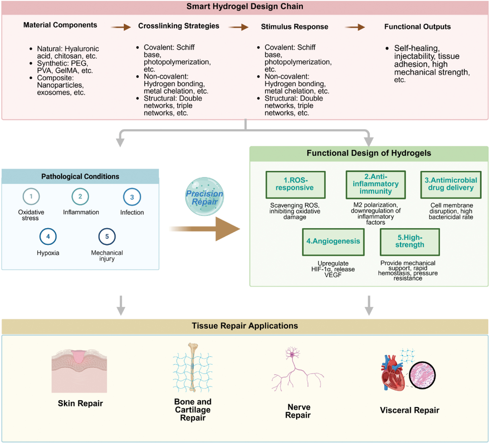
Notably, hydrogels not only act by physical occlusion and stimulation of blood coagulation factor aggregation during hemostasis, but also effectively prevent wound infections and increase repair efficiency through their inherent antimicrobial components or the antimicrobial agents they contain. ES gel, for example, has inherent antimicrobial activity that inhibits bacterial growth during hemostasis; therefore, this material is suitable for the repair of infected wounds [131]. Its ability to work without the added burden of antimicrobial agents also decreases the risk of developing drug resistance associated with overuse and confers important clinical value. Non-covalent CT hydrogels maintain excellent adhesion in moist environments, because of the natural coagulating properties of TA and the biocompatibility of collagen; these materials are therefore suitable for rapid control of visceral bleeding [140]. Notably, they meet the stringent requirements for adhesion strength and biosafety in the repair of internal organs. Figure 3 illustrates the overall framework of smart hydrogels, including material design, pathological response, and tissue repair applications.
Figure 3 Theoretical framework for the full process of smart hydrogel systems: adaptation to pathological microenvironments and tissue repair. This diagram systematically outlines the composition of smart hydrogel materials, cross-linking strategies, stimulus-responsive properties, and the design logic for functional output. It displays the mechanisms underlying the precise adaptation of hydrogels to pathological microenvironments, such as oxidative stress and inflammation, and demonstrates their applications in multi-tissue repair, thereby presenting a comprehensive end-to-end process encompassing material design, pathological adaptation, and targeted repair. Created in BioRender. Siqi, S. (2026) https://BioRender.com/0jfd3tt.
Modulating the pathological microenvironment
When pathological changes occur in organs, the pathological microenvironment of the organism changes because of damage to internal organs. However, not all changes in the microenvironment positively influence tissue regeneration. Creating a pathological microenvironment that promotes the regeneration of internal organs is crucial in the treatment of internal organ diseases. Therefore, hydrogels, which are new biomaterials that can be used to modify the pathological microenvironment of the body, are highly important in clinical treatment research. Zhang’s team has developed a new three-dimensional, biocompatible porous hydrogel (DFO gel) that mimics hypoxic conditions, increases the expression of HIF-1α and vascular endothelial growth factor (VEGF), and thus effectively repairs kidney damage [144]. Jiang et al. have developed an MnO2-based mesoporous PD(HA) hydrogel that degrades in the oxidative microenvironment of the liver and releases Mn2+ and exosomal miR-582-5p simultaneously, thus alleviating the pathological oxidative microenvironment in the liver, modulating liver-heart axis signaling, and ultimately ameliorating lipid accumulation in hepatocytes [145]. Composite hydrogels detect pathological signals, and regulate or release regenerative substances by mimicking and improving the pathological microenvironment of internal organs to achieve a regenerative effect.
Therefore, smart hydrogels stand out from other biomaterials as novel tissue regeneration materials because of their fundamental advantages (precise reaction mechanisms, versatile manufacturing techniques, and excellent biocompatibility). The development of hydrogel products has become an essential bridge connecting fundamental research with clinical applications. The immense value of these materials manifests primarily in three aspects. First, precise and controllable reaction mechanisms enable hydrogels to respond to multiple stimuli such as temperature, light, and pH, thereby enabling “on-demand” drug release. This capability significantly enhances treatment precision while eliminating delays and other limitations inherent in traditional therapies. Second, universal and highly efficient drug delivery capabilities enable hydrogels to flexibly integrate drugs, bioactive substances, and functional cells to address regenerative needs as diverse as skin lesion repair, osteochondral defect treatment, nerve regeneration, visceral hemostasis, and targeted tumor therapy. After reaching injury sites, hydrogel dressings enable comprehensive coverage from localized intervention to systemic regulation. Third, exceptional biocompatibility enables hydrogels to readily integrate with innovative technologies such as 3D bioprinting and microfluidics in the construction of biomimetic repair structures. Primarily composed of natural polymers such as collagen and HA, these materials exhibit low immunogenicity and outstanding biocompatibility. These hydrogels mimic the microenvironment of the ECM and provide stable support for tissue regeneration.
Because of these properties, smart hydrogels can overcome many limitations of traditional treatment methods, and offer more comprehensive solutions to clinical tasks such as complex wound healing, treatment of degenerative diseases, and minimally invasive treatment methods. In addition, they are practically irreplaceable. The functionalities of hydrogel materials are summarized in Table 1.
Table 1 Repair Categories, Application Scenarios, and Functional Hydrogel Materials used in Various Tissue Engineering Approaches
| Repair Type | Application Scenarios | Key Components in Relevant Research | Functionality |
|---|---|---|---|
| Skin repair | Burns | Curcumin, magnesium ions, etc. | Antibacterial, antioxidant, modulation of inflammatory responses, promotion of vascular proliferation, etc. |
| Chronic wounds | Schiff base, etc. | ||
| Acute injuries | Polylysine, AGN, etc. | ||
| Scarless repair | MSC, HA, citric acid, polyethylene glycol, AS, etc. | ||
| Bone and cartilage tissue repair | Irregular bone defects | Barium titanate (BaTiO3, BTO) nanoparticles (KBTO), biopolymers, etc. | Promotion of osteogenesis, enhancement of vascular regeneration, immune regulation, biocompatibility, degradability, etc. |
| Diabetic bone defects | TK, NB, PEG, etc. | ||
| Osteoporotic bone defects | Ca2+, SA, etc. | ||
| Osteoarthritis | Tannic acid, KGN, etc. | ||
| Osteosarcoma | Nanoparticles, etc. | ||
| Neural tissue repair | Central nervous system injury repair | HBPAK, GelMA | Simulation of the cellular growth microenvironment, matching the mechanical properties of the spinal cord, specific responsiveness, anti-inflammatory activity, mimicking the extracellular matrix within the brain, facilitating neurovascular unit reconstruction, providing biocompatible scaffolding, etc. |
| Intervention for central nervous system degenerative diseases | CMCS, F127-CHO | ||
| Peripheral nerve repair | AAm | ||
| Liver repair | Partial hepatic resection | ES gel, GelMA/OD/borax | Antibacterial and hemostatic, adhesiveness, anti-infection, biocompatibility, etc. |
| Acute liver failure | Heterogeneous microcolloid-hydrogel hybrids | ||
| Cirrhosis | Self-healing chitosan-phenol hydrogel | ||
| Kidney repair | Partial nephrectomy | Injectable HA hydrogel | Delivery cells, cell vector, self-healing properties, injectability, biocompatibility, etc. |
| Immunoglobulin nephropathy | CHO-HA/Gel-NH2 hydrogel | ||
| Kidney transplant | IGE-Gel | ||
| Heart repair | Myocardial infarction | Collagen and tannic acid, catechol-chitosan | Promotion of cell adhesion, migration, and proliferation Enhancement of mucosal adhesion and hemostatic properties, antioxidant properties, etc. |
| Myocardial ischemia | Oxidized hyaluronic acid |
Existing challenges and improvement strategies for hydrogels in tissue engineering repair
The key challenges and corresponding improvement strategies of hydrogels in tissue engineering repair are schematically illustrated in Figure 4. Hydrogels are a new biomaterial with great potential in tissue regeneration and clinical diagnostics. However, several problems and obstacles must be overcome before they can be used on a large scale. First, their properties are not sufficiently adapted to physiological requirements. Biodegradable hydrogels create a biological environment conducive to cell proliferation and tissue growth in the human body. Nicodemus and other scientists have shown that physiological stress conditions and bioreactor parameters affect their degradation [146]. This mismatch between the rate of degradation and tissue healing not only undermines the primary function of hydrogels as tissue regeneration scaffolds but also prompts safety concerns due to the instability of regeneration outcomes, thus limiting their reliable clinical use. To adapt to tissue healing, reactive enzymatic mechanisms can be used in hydrogels. Dong et al. have developed a multifunctional hydrogel dressing based on the self-organization of electrostatically induced peptides (Pep-1) and water-soluble neomethylene blue, which has rapid hemostatic effects and antibacterial properties, and accelerates wound healing [147]. By including a reactive enzymatic mechanism, the degradation rate can be dynamically adapted to the tissue healing rate. Shur et al. have found that the rate of swelling regression of PAAm hydrogels is significantly decreased when brain is used as the solvent source [148]. Compared with the initial swelling ability of hydrogels manufactured and swollen at equilibrium, the swelling ability of the same hydrogel after the dehydration cycle and implantation in brain tissue decreases by nearly 90%, thus severely limiting their long-term use in nerve tissue engineering. Because hydrogels’ water retention capacity varies depending on the temperature and solvent, strategies such as adding impermeable coatings to their surfaces can improve their stability. For example, Bai et al. have developed an adaptive lipid bilayer coating to improve water retention in hydrogel sensors [149]. This coating prevents hydrogel evaporation under static and dynamic conditions, and is non-cytotoxic.
Figure 4 Current challenges and improvement strategies for hydrogels in tissue engineering repair. The left semicircle summarizes the key bottlenecks restricting the clinical translation of hydrogels, including poor physiological adaptability, insufficient mechanical properties, single function, difficult scaling of manufacturing processes, and low design efficiency. The right semicircle presents targeted improvement strategies to address these challenges, such as dynamic regulation via enzyme control, mechanical enhancement via crosslinking, functional improvement via active component loading, cost reduction via quality control, and AI-assisted smart screening. The central schematic illustrates the application of hydrogel scaffolds in tissue engineering repair, supporting cell adhesion, proliferation, and tissue regeneration. Created in BioRender. Siqi, S. (2026) https://BioRender.com/mo34udl.
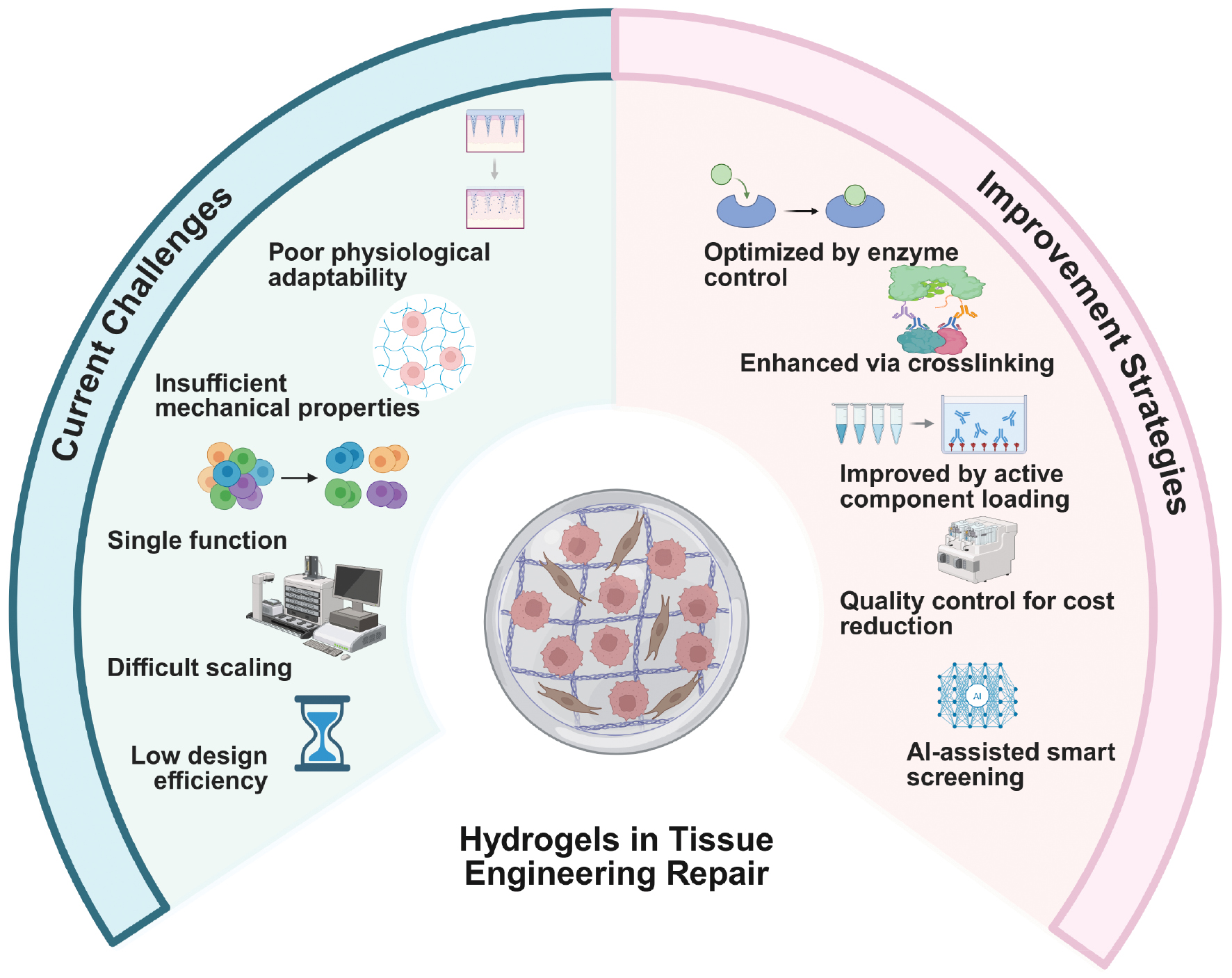
Second, a discrepancy exists between the mechanical properties and the requirements of the tissue. Most hydrogels obtained from natural compounds are characterized by low mechanical strength that cannot easily meet the requirements for supporting the growth of various tissues in the body and that prevents the creation of a stable mechanical environment. Because of the high collagen content in natural cartilage tissue, gelatin-based bioinks are particularly important for cartilage tissue engineering. However, they encounter problems such as low mechanical stability and rapid dissolution at physiological temperatures. Boretti’s team has concluded that this problem cannot be solved through a single technique [150]. To improve structural stability and cell viability, a dual cross-linking technique combining photo-induced and ionic processes is necessary. Similarly, even 3D-printed GelMA hydrogels have inevitable mechanical limitations. To overcome these limitations, the mechanical properties must be modified at several levels (monomer structure, micrometric pore design, or composite phases reinforced with nanomaterials) to meet the biomechanical requirements of soft tissue engineering. By incorporating nanomaterials into traditional hydrogels, composite systems can be created with improved mechanical strength and hardness. Carrasco’s team has incorporated reduced graphene oxide (rGO) and TA into alginate hydrogels and observed improvements in tear strength, elastic modulus, and hardness [151], thus overcoming the mechanical limitations of traditional hydrogels and achieving the strength required for in vivo biomaterials.
In addition, hydrogels have functional limitations. Classical hydrogels are usually used only as carriers or vectors, and they cannot easily meet various reconstruction requirements simultaneously, such as cell adhesion, nutrient transport, inflammatory regulation, and drug delivery. Therefore, their effectiveness and applicability in treating complex wounds, such as infected areas and chronic wounds, are substantially limited. Slow wound healing is a common clinical complication of diabetes. Traditional hydrogels cannot easily respond to stimuli, and the release of biologically active molecules in time and space is difficult to control. However, because of its protease-sensitive structure, multifunctional complex hydrogel can achieve anti-inflammatory, antioxidant, and therapeutic effects simultaneously [152]. In skin wounds, traditional hydrogel cannot meet various healing requirements, such as stopping bleeding and retaining moisture. To overcome the limitations of single functions, innovations are needed, such as iodine-encapsulated complexes and functional molecular compounds [153]. The integration of functional cells, bioactive molecules, and hydrogel matrix creates an integrated synergistic system. The use of hydrogel to provide stable structural support provides dual benefits of tissue regeneration and targeted treatment, thus overcoming the functional limitations and limited application prospects of hydrogels. Maity et al. developed a composite hydrogel that combines the hypolipidemic properties of silk fibroin (SF), the antioxidant properties of melanin, and the therapeutic effects of berberine, which can accelerate wound healing in diabetic patients [154]. The components of complex hydrogel are biocompatible, stimulate effective fibroblast migration, and control oxidative stress in vitro. This progress has transformed hydrogel from a simple matrix to a multifunctional platform capable of promoting cell regeneration and achieving targeted treatment. Furthermore, the still immature large-scale manufacturing process for hydrogels has become a major obstacle to their clinical application. The laboratory production of hydrogels often relies on manual labor or small precision instruments, and the process parameters cannot easily be scaled up correctly. Simultaneously, achieving a uniform cross-linking reaction in mass production remains a challenge, thus resulting in substantial variations among product batches in key metrics such as mechanical properties, degradation rates, and functional activity. This variability does not meet the strict stability requirements for clinical applications. Furthermore, manufacturing complex smart hydrogels (such as multifunctional composite hydrogels or stimulation-responsive hydrogels) requires complicated processes such as precise mixing of multiple components and stepwise cross-linking. Because existing production facilities lack effective automation capabilities, low production efficiency and high costs hinder large-scale delivery. Furthermore, the lack of mature solutions to challenges such as raw material loss and contamination control during manufacturing further hinders the transition of hydrogels from laboratory samples to routine clinical products. To overcome these challenges, technology-driven innovation of the manufacturing process is required: the development of automated continuous production systems that use intelligent sensor technology to dynamically control critical parameters such as temperature, pH, and cross-linking concentration, would ensure process stability in large-scale manufacturing. Precision manufacturing techniques such as microfluidic chips and 3D-printed arrays should be used to accurately replicate the microstructure of hydrogels while enabling scalable production, thereby optimizing the manufacturing workflow. A comprehensive quality management system must be established that can perform multi-point checks on the purity of raw materials, the performance of intermediate products, and the specifications of final products to minimize variations across batches. Simultaneously, low-cost alternatives with high purity should be developed, and cleaning and modification processes should be simplified to decrease energy consumption and production costs. These initiatives would lay a technical foundation for the large-scale clinical application of hydrogels. Finally, various problems persist in the selection and development of hydrogel materials. The process is deeply complex. The suitable type and quantity of building blocks must be selected among tens of thousands of components to construct hydrogel materials; in addition, the internal structures of these components introduce diversity and uncertainty. Moreover, during the actual design process, the effects of the specific environment in which the hydrogel will be applied must be considered. This aspect is important, because it can change many parts of the hydrogel. Furthermore, the current process for screening and designing hydrogels still relies on the traditional experimental trial-and-error method. This method is widely used, but it has drawbacks. It requires considerable human and material resources and cannot accurately account for the numerous variable factors in the material design process. Consequently, the screening and design of hydrogels is difficult, and progress has been substantially hindered. The introduction of AI-powered intelligent hydrogel design methods provides new possibilities for addressing these difficulties. This method uses ML, also combined with data mining and other technologies. It builds a very large 3D space by permutation and combination and then, through algorithm analysis, filters out the unreasonable theoretical models in a stepwise manner. Compared with the traditional experimental trial-and-error method, it can enable substantial human and material resource savings. Simultaneously, its high precision and efficiency make it suitable for real-world design work. However, current AI-assisted methods for screening and designing hydrogels still lack standardized data to describe various physical and chemical parameters that influence hydrogel properties. The screening process still depends on the accidental discovery of amino acid sequences, such as those from natural proteins and experimental findings. Therefore, Zhang’s team has used a combination of ML and coarse-grained molecular dynamics (CGMD) to iteratively interact with a natural tetrapeptide hydrogel library [155]. They have established a dedicated scoring function to evaluate the gelation abilities of 160,000 natural tetrapeptides, thus providing standardized data for AI-based hydrogel screening and predicting the ability of tetrapeptides to form hydrogels. The development of hydrogels by using AI has undergone a revolutionary shift from passive discovery to active research. This approach has immense potential for advancement and offers new solutions across diverse fields such as biomedicine and industrial rehabilitation.
The road to routine use of hydrogels in the medical field remains fraught with obstacles. The greatest challenges include the mismatch between the properties of the materials and physiological requirements, the differences between mechanical strength and tissue strength, limitations due to functional specificity, the still immature mass production processes, and the complexity of material selection and development. These problems highlight the conceptual shortcomings of the materials and reveal a significant incompatibility with the complex physiological environment of the human body and the various requirements for regeneration. This critical shortcoming requires an immediate solution. To fully exploit the potential of hydrogels as new biomaterials and close the gap between laboratory research and clinical application, concrete solutions to these interrelated material challenges must be identified to achieve multifunctional optimization.
Future development directions and clinical translation expectations for hydrogels
Future development of hydrogels, innovative biomaterials with biocompatibility and functional plasticity, should focus not only on integrated multifunctional applications but also on specific clinical problems. Figure 5 presents the practical problems in the current development of intelligent hydrogels, and also provides solutions and future prospects for such hydrogels. Key objectives such as problem solving, safety improvement, and implementation into practice will require bridging the gap between laboratory prototypes and clinical products used in everyday practice. The three pillars underlying this transformation are deep integration of AI, removal of barriers between the laboratory and the clinical environment, and strict adaptation of medical needs to technical solutions. These pillars provide a sustainable dynamic for the clinical application of hydrogels.
Figure 5 Schematic diagram of future development and clinical translation of smart hydrogels. This figure illustrates the future development directions of smart hydrogels across six specific dimensions: precision and personalization; large-scale production; medical-engineering integration; terminal sterilization; clinical translation pathway; and quality and standardization. These correspond to the following six development directions: data-driven personalized hydrogel design, low-cost large-scale production, medical-production engineering integrated R&D, low-temperature sterilization, multidisciplinary collaboration for translation, and establishment of a standard supervision system. Created in BioRender. q&, lÉ. (2026) https://BioRender.com/2hybnau.
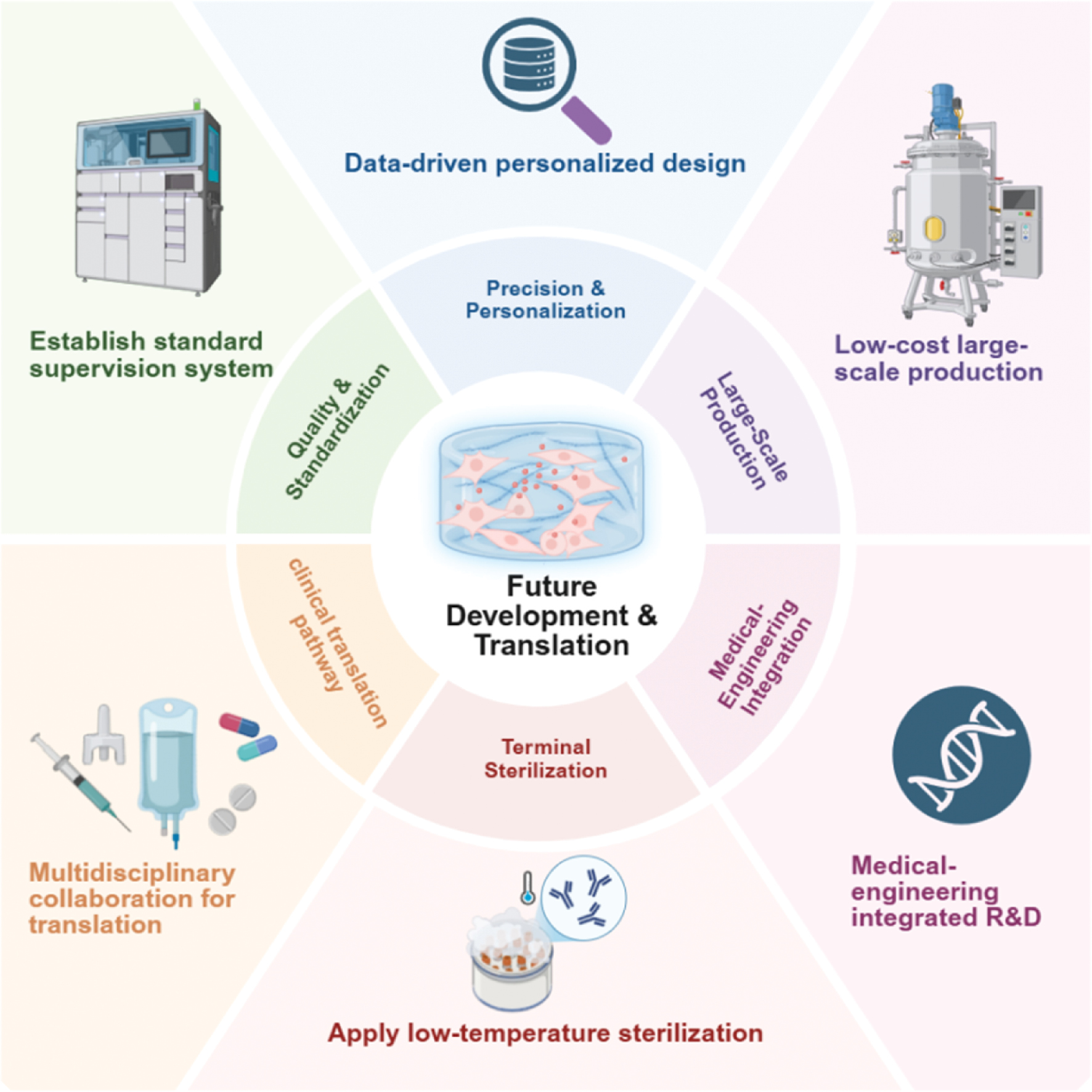
A standardized quality assurance system must be implemented, characterized by clear classification, comprehensive testing, and appropriate regulations. First, clear criteria must be established to define the properties of hydrogel-based products. Hydrogels should be classified as drugs or medical devices, depending on their primary function, and appropriate regulatory procedures should be applied. Next, a comprehensive testing system covering the entire product life cycle must be implemented. In addition to traditional biocompatibility and degradability, important indicators such as functional stability (e.g., sensitivity to reaction after long-term storage) and in vivo metabolic safety (e.g., assessment of the toxicity of degradation products to organs) must be considered. Standardized testing decreases clinical risks and strengthens safety barriers in clinical applications.
From a technical standpoint, data-driven precision engineering will form the basis for personalized therapies. By combining the mechanical properties of the patient’s tissues and pathological parameters of the microenvironment (e.g., pH, enzymatic activity, and inflammatory factors) with a database of materials (cross-linking methods, component proportions, and microstructural parameters), highly effective personalized hydrogels can be obtained. For example, in the case of damage to the cartilage that supports body weight, fine-tuning the elasticity and compression resistance coefficients to match natural cartilage improves both mechanical support during repair and tissue regeneration. This design paradigm will gradually replace the traditional trial-and-error approach and establish a standard workflow comprising clinical requirements → data modeling → performance optimization → verification and iteration. Because of this development, the functionality of hydrogels is expected to change from general adaptation to precise and personalized adaptation, thus providing the necessary material basis for personalized therapies. In parallel, data collected during long-term clinical observation are expected to provide additional information on the metabolic pathways of hydrogel degradation in the body, their long-term biological safety, and functional degradation patterns, thus providing continuous clinical feedback that can be used for optimization.
The introduction of a large-scale, cost-effective, and highly stable manufacturing process is a crucial prerequisite for the clinical application of hydrogels. Currently, the manufacture of smart hydrogels faces challenges such as substantial variations across batches, high costs, and difficulties in scaling up production. Future solutions will require process innovation. First, precision manufacturing techniques must accurately reproduce the microstructure of hydrogels and enable continuous production. Simultaneously, intelligent control systems must adjust critical parameters such as preparation temperature and cross-linking time to ensure consistent performance across all batches. Simultaneously, optimization of the raw material system through the development of inexpensive, high-purity, naturally derived substances is necessary to simplify the preparation process and decrease material waste.
The integration of medicine and technology is accelerating the clinical use of hydrogels. Clinicians involved in research and development ensure that innovations in materials can work in real clinical scenarios. For chronic wounds with recurrent infections and delayed healing, composite hydrogels may be developed that combine long-term antimicrobial release with inflammation regulation. For joint cartilage regeneration, functional hydrogels may be developed that provide excellent mechanical support and rapid tissue integration. For diabetic foot ulcers requiring nerve regeneration, tactile adaptation capabilities may be integrated. This closed collaboration model (involving identifying clinical needs, developing technical solutions, and confirming clinical efficacy) effectively bridges the gap between research and development and practical application. Consequently, hydrogel-based products can be transformed from functional prototypes limited to laboratory environments into truly effective clinical tools.
Innovations in terminal sterilization technology eliminate the final barrier to the clinical application of hydrogels. Conventional high-temperature, high-pressure sterilization methods often damage the bioactive components and reactive structures of hydrogels. Therefore, low-temperature sterilization technology is used for the manufacture of smart hydrogels. This approach preserves the basic structure of the hydrogel and maintains its smart reactive properties without damage. The mature application of low-temperature sterilization technology completely solves the industry’s challenge of the contradiction between sterilization and activity preservation, and provides technical assurance for the use of hydrogels in clinical environments such as operating rooms and wound treatment centers.
Therefore, the transition of hydrogels from functionalization to clinical effectiveness will require multidimensional and systematic breakthrough innovations. Research is expected to continue to drive the development of hydrogels combining intelligent responsiveness, mechanical adaptability, and functional integration. This aim is expected to be achieved by integrating knowledge from materials science, biology, clinical medicine, and production technology; establishing a standardized quality system as the foundation; focusing on precision design; using the integration of medicine and technology as a fundamental principle; supporting large-scale production; and ensuring safe sterilization. These advancements would enable the substantial potential of hydrogels to be fully realized in areas such as tissue reconstruction, regenerative medicine, and the treatment of chronic diseases, and establish hydrogels as an important biomaterial that stimulates innovation in clinical treatment models.
Acknowledgement
This study was supported by the Natural Science Foundation of Heilongjiang Province (No. LH2024H013) and State Key Laboratory of Frigid Zone Cardiovascular Diseases (SKLFZCD) Open Project (No. HDHY2025024). The Graphical Abstract was created in BioRender. S, B. (2026) https://BioRender.com/etstzqb.
Conflict of interest
The authors declare that there are no conflicts of interest.
Graphical abstract
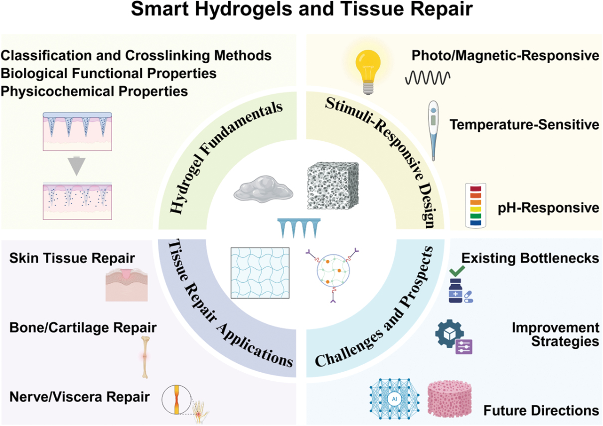
Highlights
- Clinical challenges in tissue repair push smart hydrogels to forefront research in this field.
- Smart hydrogels are classified by stimulus responsiveness, with unique physicochemical properties enabling dynamic adaptation to repair requirements.
- Through multiple mechanisms, hydrogels demonstrate effective repair capabilities across diverse tissue repair domains.
- This paper systematically reviews recent advancements and explores the immense potential of smart hydrogels in tissue repair.
- The research provides robust support for addressing challenges and guiding future application of smart hydrogels in tissue repair.
In brief
Tissue regeneration poses major challenges in clinical medicine, and numerous biomaterials have been shown to accelerate tissue regeneration. Smart hydrogels have attracted research attention in tissue regeneration, because of their exceptional physical-chemical and biological properties, high water content, excellent biocompatibility, flexibility, degradability, and ability to respond to various external stimuli. This article focuses on smart hydrogels’ enormous potential for tissue regeneration and their classification. It describes their areas of application and mechanisms of action in skin, bone and cartilage, nerve, and visceral tissue regeneration. Current issues and strategies for improving hydrogels in tissue regeneration through tissue engineering are examined. Finally, the article highlights the intelligent alignment of advances through full integration into the field of artificial intelligence, which may provide a reference for research in related fields.
References
- Guo P, Lei P, Luo L, Yang Q, Yang Q, et al. Microfluidic-engineered Chinese herbal nanocomposite hydrogel microspheres for diabetic wound tissue regeneration. J Nanobiotechnol 2024;22(1):724. [PMID: 39568066 DOI: 10.1186/s12951-024-02998-0]
- Zhao Z, Zhang Y, Meng C, Xie X, Cui W, et al. Tissue-penetrating ultrasound-triggered hydrogel for promoting microvascular network reconstruction. Adv Sci (Weinh) 2024;11(23):e2401368. [PMID: 38600702 DOI: 10.1002/advs.202401368]
- Peña OA, Martin P. Cellular and molecular mechanisms of skin wound healing. Nat Rev Mol Cell Biol 2024;25(8):599-616. [PMID: 38528155 DOI: 10.1038/s41580-024-00715-1]
- Nie R, Zhang QY, Feng ZY, Huang K, Zou CY, et al. Hydrogel-based immunoregulation of macrophages for tissue repair and regeneration. Int J Biol Macromol 2024;268(Pt 1):131643. [PMID: 38643918 DOI: 10.1016/j.ijbiomac.2024.131643]
- Nii T, Katayama Y. Biomaterial-assisted regenerative medicine. Int J Mol Sci 2021;22(16):8657. [PMID: 34445363 DOI: 10.3390/ijms22168657]
- Maurya PK, Hazarika K, Bandyopadhyay-Ghosh S, Nandi SK, Ghosh SB. From water to medicine: the transformative role of hydrogels in diabetic wound healing. J Biomater Sci Polym Ed 2025;1-26. [PMID: 41359509 DOI: 10.1080/09205063.2025.2599294]
- Qing X, He G, Liu Z, Yin Y, Cai W, et al. Preparation and properties of polyvinyl alcohol/N–succinyl chitosan/lincomycin composite antibacterial hydrogels for wound dressing. Carbohydr Polym 2021;261:117875. [PMID: 33766362 DOI: 10.1016/j.carbpol.2021.117875]
- Hao R, Cui Z, Zhang X, Tian M, Zhang L, et al. Rational design and preparation of functional hydrogels for skin wound healing. Front Chem 2022;9:839055. [PMID: 35141209 DOI: 10.3389/fchem.2021.839055]
- Delanne-Cumémal A, Verney V, Portanguen S, Fadhlaoui K, Chalancon S, et al. Polysaccharides from Adansonia digitata combined with whey protein and alginate enhance the viscosity, swelling, controlled release, and mucoadhesion properties of hydrogels for oral drug delivery. Int J Pharm 2026;692:126624. [PMID: 41592628 DOI: 10.1016/j.ijpharm.2026.126624]
- Pavithra B, Singh P, Ramesh Kumar V, Durairaj S, Hassan S. Advances in polymeric nanoparticles and hydrogels in 3D bioprinting: enhancing bioinks for tissue engineering and regenerative medicine. Bioprinting 2025;51:e00438. [DOI: 10.1016/j.bprint.2025.e00438]
- Shen F, Ding X, Wang G, Chen M, Shi H, et al. Dextran based hydrogel wound dressing with cytocompatible, anti-protein and antibacterial properties for infected wound healing. Int J Biol Macromol 2025;334(Pt 1):148709. [PMID: 41207592 DOI: 10.1016/j.ijbiomac.2025.148709]
- Hamidi M, Azadi A, Rafiei P. Hydrogel nanoparticles in drug delivery. Adv Drug Deliv Rev 2008;60(15):1638-49. [PMID: 18840488 DOI: 10.1016/j.addr.2008.08.002]
- Kass LE, Nguyen J. Nanocarrier-hydrogel composite delivery systems for precision drug release. Wiley Interdiscip Rev Nanomed Nanobiotechnol 2022;14(2):e1756. [PMID: 34532989 DOI: 10.1002/wnan.1756]
- He Q, Ding X, Deng J, Zhang Y, Wang X, et al. Fabrication of injectable, adhesive, self-healing, superabsorbent hydrogels based on quaternary ammonium chitosan and oxidized pullulan. Heliyon 2024;10(19):e38577. [PMID: 39435091 DOI: 10.1016/j.heliyon.2024.e38577]
- Rafati F, Johari N, Nokhbedehghan Z, Samadikuchaksaraei A, Razavi M, et al. Innovative silk fibroin-flax nanocomposite hydrogels: a drug-loaded system for potential wound dressing applications. Mater Today Commun 2025;49:113973. [DOI: 10.1016/j.mtcomm.2025.113973]
- Yu H, Zhang J, Yang L, Tian Y, Milne C, et al. MSC-derived exosomes injectable hyaluronic acid hydrogel for enhanced chronic wound healing. J Control Release 2025;385:113985. [PMID: 40581219 DOI: 10.1016/j.jconrel.2025.113985]
- Hoffman AS. Hydrogels for biomedical applications. Adv Drug Deliv Rev 2002;54(1):3-12. [PMID: 11755703 DOI: 10.1016/s0169-409x(01)00239-3]
- Arguchinskaya NV, Isaeva EV, Kisel AA, Beketov EE, Lagoda TS, et al. Properties and printability of the synthesized hydrogel based on GelMA. Int J Mol Sci 2023;24(3):2121. [PMID: 36768446 DOI: 10.3390/ijms24032121]
- Duan Y, Jiang F, Li Q, McDowell A, Li Y, et al. Multifunctional polysaccharide/metal/polyphenol double-crosslinked hydrogel for infected wound. Carbohydr Polym 2024;332:121912. [PMID: 38431415 DOI: 10.1016/j.carbpol.2024.121912]
- Zhao J, Jia F, Li J, Tao YC, Hu JY, et al. Sprayable reactive oxygen species-responsive hydrogel coatings restore endothelial barrier integrity for functional vascular healing. ACS Nano 2025;19(23):21757-74. [PMID: 40457543 DOI: 10.1021/acsnano.5c05477]
- Lin X, Liu R, Zhou Y, Beitzel J, Noguchi A, et al. Injectable biodegradable polysaccharide-based hydrogels for stem cell delivery and cartilage regeneration. J Mater Chem B 2025;13(26):7838-53. [PMID: 40485218 DOI: 10.1039/d5tb00287g]
- Wang Z, Liu J, Deng R, Yang Y, Fangfang C, et al. Bioactive and biodegradable hydrogel based on the dermal matrix loaded with microspheres containing vascular endothelial growth factor (VEGF) and interleukin (IL)-10 accelerated the healing of diabetic wounds in rats. Regen Ther 2025;29:575-86. [PMID: 40475694 DOI: 10.1016/j.reth.2025.04.017]
- Guo L, Fu Z, Li H, Wei R, Guo J, et al. Smart hydrogel: a new platform for cancer therapy. Adv Colloid Interface Sci 2025;340:103470. [PMID: 40086017 DOI: 10.1016/j.cis.2025.103470]
- Jiang Y, Zhu C, Ma X, Fan D. Smart hydrogel-based trends in future tendon injury repair: a review. Int J Biol Macromol 2024;282(Pt 5):137092. [PMID: 39489238 DOI: 10.1016/j.ijbiomac.2024.137092]
- Cai J, Guo J, Wang S. Application of polymer hydrogels in the prevention of postoperative adhesion: a review. Gels 2023;9(2):98. [PMID: 36826268 DOI: 10.3390/gels9020098]
- Ghanbari M, Salavati-Niasari M, Mohandes F, Firouzi Z. Modified silicon carbide NPs reinforced nanocomposite hydrogels based on alginate-gelatin by with high mechanical properties for tissue engineering. Arab J Chem 2022;15(1):103520. [DOI: 10.1016/j.arabjc.2021.103520]
- Zhang X, Liang Y, Huang S, Guo B. Chitosan-based self-healing hydrogel dressing for wound healing. Adv Colloid Interface Sci 2024;332:103267. [PMID: 39121832 DOI: 10.1016/j.cis.2024.103267]
- He J, Sun Y, Gao Q, He C, Yao K, et al. Gelatin methacryloyl hydrogel, from standardization, performance, to biomedical application. Adv Healthc Mater 2023;12(23):e2300395. [PMID: 37115708 DOI: 10.1002/adhm.202300395]
- Hong SB, Jeong JO, Choi H. Hydrogels in cardiac surgery: versatile platforms for tissue repair, adhesion prevention, and localized therapeutics. Gels 2025;11(7):564. [PMID: 40710725 DOI: 10.3390/gels11070564]
- Dobner S, Bezuidenhout D, Govender P, Zilla P, Davies N. A synthetic non-degradable polyethylene glycol hydrogel retards adverse post-infarct left ventricular remodeling. J Card Fail 2009;15(7):629-36. [PMID: 19700140 DOI: 10.1016/j.cardfail.2009.03.003]
- Gong D, Wu N, Chen H, Zhang W, Yan C, et al. Phytic acid-loaded polyvinyl alcohol hydrogel promotes wound healing of injured corneal epithelium through inhibiting ferroptosis. Redox Biol 2024;76:103354. [PMID: 39298836 DOI: 10.1016/j.redox.2024.103354]
- Visan AI, Negut I. Sustainable hydrogels in water treatment—a short review. Gels 2025;11(10):812. [PMID: 41149418 DOI: 10.3390/gels11100812]
- Parvin N, Kumar V, Joo SW, Mandal TK. Cutting-edge hydrogel technologies in tissue engineering and biosensing: an updated review. Materials (Basel) 2024;17(19):4792. [PMID: 39410363 DOI: 10.3390/ma17194792]
- Segneanu AE, Bejenaru LE, Bejenaru C, Blendea A, Mogoşanu GD, et al. Advancements in hydrogels: a comprehensive review of natural and synthetic innovations for biomedical applications. Polymers (Basel) 2025;17(15):2026. [PMID: 40808075 DOI: 10.3390/polym17152026]
- Liu Z, Ma X, Liu J, Zhang H, Fu D. Advances in the application of natural/synthetic hybrid hydrogels in tissue engineering and delivery systems: a comprehensive review. Int J Pharm 2025;672:125323. [PMID: 39923883 DOI: 10.1016/j.ijpharm.2025.125323]
- Bustamante-Torres M, Romero-Fierro D, Arcentales-Vera B, Palomino K, Magana H, et al. Hydrogels classification according to the physical or chemical interactions and as stimuli-sensitive materials. Gels 2021;7(4):182. [PMID: 34842654 DOI: 10.3390/gels7040182]
- Potaś J, Szymańska E, Winnicka K. Challenges in developing of chitosan—based polyelectrolyte complexes as a platform for mucosal and skin drug delivery. Eur Polym J 2020;140:110020. [DOI: 10.1016/j.eurpolymj.2020.110020]
- Li H, Peng M, Li J, Wang L, Do H, et al. SO2F2 mediated click chemistry enables modular disulfide formation in diverse reaction media. Nat Commun 2024;15(1):8325. [PMID: 39333088 DOI: 10.1038/s41467-024-52606-w]
- Kim SH, Kim K, Kim BS, An YH, Lee UJ, et al. Fabrication of polyphenol-incorporated anti-inflammatory hydrogel via high-affinity enzymatic crosslinking for wet tissue adhesion. Biomaterials 2020;242:119905. [PMID: 32145505 DOI: 10.1016/j.biomaterials.2020.119905]
- Wei L, Yang Y, Qiu X, Shen J, Zhao Y, et al. Self-polymerized tough and high-entanglement zwitterionic functional hydrogels. Small 2024;20(50):e2405789. [PMID: 39319480 DOI: 10.1002/smll.202405789]
- Feng Q, Zhang M, Zhang G, Mei H, Su C, et al. A whole-course-repair system based on ROS/glucose stimuli-responsive EGCG release and tunable mechanical property for efficient treatment of chronic periodontitis in diabetic rats. J Mater Chem B 2024;12(15):3719-40. [PMID: 38529844 DOI: 10.1039/d3tb02898d]
- Li X, Xiong Y. Application of “click” chemistry in biomedical hydrogels. ACS Omega 2022;7(42):36918-28. [PMID: 36312409 DOI: 10.1021/acsomega.2c03931]
- Zhang Z, Tao ZZ, Du R, Huo R, Zheng X. Artificial intelligence informed hydrogel biomaterials in additive manufacturing. Gels 2025;11(12):981. [PMID: 41441137 DOI: 10.3390/gels11120981]
- Sokmen S, Cakmak S, Oksuz I. 3D printing of an artificial intelligence-generated patient-specific coronary artery segmentation in a support bath. Biomed Mater 2024;19(3):035038. [PMID: 38626778 DOI: 10.1088/1748-605X/ad3f60]
- Li Z, Song P, Li G, Han Y, Ren X, et al. AI energized hydrogel design, optimization and application in biomedicine. Mater Today Bio 2024;25:101014. [PMID: 38464497 DOI: 10.1016/j.mtbio.2024.101014]
- Hill H, Roadevin C, Duffy S, Mandrik O, Brentnall A. Cost-effectiveness of AI for risk-stratified breast cancer screening. JAMA Netw Open 2024;7(9):e2431715. [PMID: 39235813 DOI: 10.1001/jamanetworkopen.2024.31715]
- Yu Y, Liang X, Ruan H, Wang T, Li Y, et al. Hydrogel-based sensors for multimodal health monitoring: from material design to intelligent sensing. Nanoscale 2025;17(43):24805-29. [PMID: 41144230 DOI: 10.1039/d5nr03553h]
- Liao H, Hu S, Yang H, Wang L, Tanaka S, et al. Data-driven de novo design of super-adhesive hydrogels. Nature 2025;644(8075):89-95. [PMID: 40770436 DOI: 10.1038/s41586-025-09269-4]
- Salehi S, Naghib SM, Garshasbi HR, Ghorbanzadeh S, Zhang W. Smart stimuli-responsive injectable gels and hydrogels for drug delivery and tissue engineering applications: a review. Front Bioeng Biotechnol 2023;11:1104126. [PMID: 36911200 DOI: 10.3389/fbioe.2023.1104126]
- Cai Y, Xin L, Sun P, Li H, Liu C, et al. Temperature-sensitive multifunctional intelligent responsive hydrogel based on carboxymethyl agarose and N-isopropylacrylamide: controlled drug release and accelerated wound healing. Carbohydr Polym 2023;322:121327. [PMID: 37839839 DOI: 10.1016/j.carbpol.2023.121327]
- Zhang Y, Shi K, Yang X, Chen W, Wang T, et al. Sustained release of levobupivacaine from temperature-sensitive injectable hydrogel for long-term local anesthesia in postoperative pain management. Biomaterials 2023;299:122129. [PMID: 37167892 DOI: 10.1016/j.biomaterials.2023.122129]
- Gui H, Yang T, Li LL, Liang F, Yang Z. Temperature-sensitive anti-inflammatory organohydrogels containing Janus particle stabilized phase-change microinclusions. ACS Nano 2022;16(6):9859-70. [PMID: 35699249 DOI: 10.1021/acsnano.2c03940]
- Wang R, Yang Z, Luo J, Hsing IM, Sun F. B12-dependent photoresponsive protein hydrogels for controlled stem cell/protein release. Proc Natl Acad Sci U S A 2017;114(23):5912-7. [PMID: 28533376 DOI: 10.1073/pnas.1621350114]
- Wang H, Morales RT, Cui X, Huang J, Qian W, et al. A photoresponsive hyaluronan hydrogel nanocomposite for dynamic macrophage immunomodulation. Adv Healthc Mater 2019;8(4):e1801234. [PMID: 30537061 DOI: 10.1002/adhm.201801234]
- Fan L, Zhang X, Liu X, Sun B, Li L, et al. Responsive hydrogel microcarrier-integrated microneedles for versatile and controllable drug delivery. Adv Healthc Mater 2021;10(9):e2002249. [PMID: 33690992 DOI: 10.1002/adhm.202002249]
- Chen Y, Wang X, Tao S, Wang Q, Ma PQ, et al. Research advances in smart responsive-hydrogel dressings with potential clinical diabetic wound healing properties. Mil Med Res 2023;10(1):37. [PMID: 37608335 DOI: 10.1186/s40779-023-00473-9]
- Long J, Zhou G, Yu X, Xu J, Hu L, et al. Harnessing chemical functionality of xylan hemicellulose towards carbohydrate polymer-based pH/magnetic dual-responsive nanocomposite hydrogel for drug delivery. Carbohydr Polym 2024;343:122461. [PMID: 39174134 DOI: 10.1016/j.carbpol.2024.122461]
- Park SH, Shin HS, Park SN. A novel pH-responsive hydrogel based on carboxymethyl cellulose/2-hydroxyethyl acrylate for transdermal delivery of naringenin. Carbohydr Polym 2018;200:341-52. [PMID: 30177174 DOI: 10.1016/j.carbpol.2018.08.011]
- Wu Y, Wang Y, Long L, Hu C, Kong Q, et al. A spatiotemporal release platform based on pH/ROS stimuli-responsive hydrogel in wound repairing. J Control Release 2022;341:147-65. [PMID: 34813880 DOI: 10.1016/j.jconrel.2021.11.027]
- Yi M, Jin W, Li H, Niu X, Wang J, et al. pH-responsive bilayer hydrogel with sequential release of morin-based nanoparticles and bFGF for the treatment of the “ice and fire” wounds. Materials Today Bio 2025;35:102519. [PMID: 41356418 DOI: 10.1016/j.mtbio.2025.102519]
- Hia EM, Suh IW, Jang SR, Park CH. Magnetically responsive micro-clustered calcium phosphate-reinforced cell-laden microbead sodium alginate hydrogel for accelerated osteogenic tissue regeneration. Carbohydr Polym 2024;346:122666. [PMID: 39245476 DOI: 10.1016/j.carbpol.2024.122666]
- Xue B, Peng Y, Zhang Y, Yang S, Zheng Y, et al. A novel superparamagnetic-responsive hydrogel facilitates disc regeneration by orchestrating cell recruitment, proliferation, and differentiation within hostile inflammatory niche. Adv Sci (Weinh) 2024;11(44):e2408093. [PMID: 39373392 DOI: 10.1002/advs.202408093]
- Manescu Paltanea V, Dumitru AV, Antoniac A, Antoniac I, Paltanea G, et al. Magnetic hydrogels as a treatment for oncological pathologies. J Funct Biomater 2025;16(11):414. [PMID: 41295069 DOI: 10.3390/jfb16110414]
- Qiao L, Liang Y, Chen J, Huang Y, Alsareii SA, et al. Antibacterial conductive self-healing hydrogel wound dressing with dual dynamic bonds promotes infected wound healing. Bioact Mater 2023;30:129-41. [PMID: 37554541 DOI: 10.1016/j.bioactmat.2023.07.015]
- Yao WD, Zhou JN, Tang C, Zhang JL, Chen ZY, et al. Hydrogel microneedle patches loaded with stem cell mitochondria-enriched microvesicles boost the chronic wound healing. ACS Nano 2024;18(39):26733-50. [PMID: 39238258 DOI: 10.1021/acsnano.4c06921]
- He Y, Li Y, Sun Y, Zhao S, Feng M, et al. A double-network polysaccharide-based composite hydrogel for skin wound healing. Carbohydr Polym 2021;261:117870. [PMID: 33766357 DOI: 10.1016/j.carbpol.2021.117870]
- Zhang H, Zhou W, Wang H, Zhang J, Yang H, et al. Hydrogel-based bioactive synthetic skin stimulates regenerative gas signaling and eliminates interfacial pathogens to promote burn wound healing. ACS Nano 2025;19(15):15002-17. [PMID: 40205977 DOI: 10.1021/acsnano.5c01134]
- Chen S, Xiong Y, Yang F, Hu Y, Feng J, et al. Approaches to scarless burn wound healing: application of 3D printed skin substitutes with dual properties of anti-infection and balancing wound hydration levels. EBioMedicine 2024;106:105258. [PMID: 39068733 DOI: 10.1016/j.ebiom.2024.105258]
- Zhang T, Sheng S, Cai W, Yang H, Li J, et al. 3-D bioprinted human-derived skin organoids accelerate full-thickness skin defects repair. Bioact Mater 2024;42:257-69. [PMID: 39285913 DOI: 10.1016/j.bioactmat.2024.08.036]
- Tedesco DJ, Hutter MF, Khalaf F, Ricciuti Z, Jeschke MG. Sepsis in burn care: incidence and outcomes. Mil Med Res 2025;12(1):55. [PMID: 40890875 DOI: 10.1186/s40779-025-00643-x]
- Gong Y, Wang P, Cao R, Wu J, Ji H, et al. Exudate absorbing and antimicrobial hydrogel integrated with multifunctional curcumin-loaded magnesium polyphenol network for facilitating burn wound healing. ACS Nano 2023;17(22):22355-70. [PMID: 37930078 DOI: 10.1021/acsnano.3c04556]
- Sun J, Jia W, Qi H, Huo J, Liao X, et al. An antioxidative and active shrinkage hydrogel integratedly promotes re-epithelization and skin constriction for enhancing wound closure. Adv Mater 2024;36(21):e2312440. [PMID: 38332741 DOI: 10.1002/adma.202312440]
- Zhang F, Zhang H, Wang S, Gao M, Du K, et al. A dynamically phase-adaptive regulating hydrogel promotes ultrafast anti-fibrotic wound healing. Nat Commun 2025;16(1):3738. [PMID: 40254609 DOI: 10.1038/s41467-025-58987-w]
- Zhang M, Xiang C, Zhen X, Luo W, He X, et al. Natural polymer based drug-loaded hydrogel platform for comprehensive care of acute spinal cord injury. Mater Today Bio 2025;31:101464. [PMID: 39896281 DOI: 10.1016/j.mtbio.2025.101464]
- Wang S, Wang R, Chen J, Yang B, Shu J, et al. Controlled extracellular vesicles release from aminoguanidine nanoparticle-loaded polylysine hydrogel for synergistic treatment of spinal cord injury. J Control Release 2023;363:27-42. [PMID: 37722419 DOI: 10.1016/j.jconrel.2023.09.026]
- Liu M, Wang Y, Wang H, Qi L, Shang Y, et al. Electret-inspired charge-injected hydrogel for scar-free healing of bacterially infected burns through bioelectrical stimulation and immune modulation. Adv Sci (Weinh) 2025;12(13):e2411889. [PMID: 39951351 DOI: 10.1002/advs.202411889]
- Su Y, Zhang M, Yu B, Tian F, Zhu D, et al. Temperature-programmable deformable microneedles for scar-free healing of infective wounds via sensory nerve regeneration. Small 2025;21(20):e2501491. [PMID: 40178015 DOI: 10.1002/smll.202501491]
- Xiong M, Yang X, Shi Z, Xiang J, Gao H, et al. Programmable artificial skins accomplish antiscar healing with multiple appendage regeneration. Adv Mater 2024;36(50):e2407322. [PMID: 39460410 DOI: 10.1002/adma.202407322]
- Yang Y, Zhang J, Wu S, Deng Y, Wang S, et al. Exosome/antimicrobial peptide laden hydrogel wound dressings promote scarless wound healing through miR-21-5p-mediated multiple functions. Biomaterials 2024;308:122558. [PMID: 38581764 DOI: 10.1016/j.biomaterials.2024.122558]
- Zhao X, Luo J, Huang Y, Mu L, Chen J, et al. Injectable antiswelling and high-strength bioactive hydrogels with a wet adhesion and rapid gelling process to promote sutureless wound closure and scar-free repair of infectious wounds. ACS Nano 2023;17(21):22015-34. [PMID: 37862553 DOI: 10.1021/acsnano.3c08625]
- Park J, Kim TY, Kim Y, An S, Kim KS, et al. A mechanically resilient and tissue-conformable hydrogel with hemostatic and antibacterial capabilities for wound care. Adv Sci (Weinh) 2023;10(30):e2303651. [PMID: 37705116 DOI: 10.1002/advs.202303651]
- Sun H, Wang S, Yang F, Tan M, Bai L, et al. Conductive and antibacterial dual-network hydrogel for soft bioelectronics. Mater Horiz 2023;10(12):5805-21. [PMID: 37817573 DOI: 10.1039/d3mh00813d]
- Guo S, Ren Y, Chang R, He Y, Zhang D, et al. Injectable self-healing adhesive chitosan hydrogel with antioxidative, antibacterial, and hemostatic activities for rapid hemostasis and skin wound healing. ACS Appl Mater Interfaces 2022;14(30):34455-69. [PMID: 35857973 DOI: 10.1021/acsami.2c08870]
- Xu Z, Liu G, Liu P, Hu Y, Chen Y, et al. Hyaluronic acid-based glucose-responsive antioxidant hydrogel platform for enhanced diabetic wound repair. Acta Biomater 2022;147:147-57. [PMID: 35649507 DOI: 10.1016/j.actbio.2022.05.047]
- Song Y, You Y, Xu X, Lu J, Huang X, et al. Adipose-derived mesenchymal stem cell-derived exosomes biopotentiated extracellular matrix hydrogels accelerate diabetic wound healing and skin regeneration. Adv Sci (Weinh) 2023;10(30):e2304023. [PMID: 37712174 DOI: 10.1002/advs.202304023]
- Liu W, Gao R, Yang C, Feng Z, Ou-Yang W, et al. ECM-mimetic immunomodulatory hydrogel for methicillin-resistant Staphylococcus aureus–infected chronic skin wound healing. Sci Adv 2022;8(27):eabn7006. [PMID: 35857459 DOI: 10.1126/sciadv.abn7006]
- Jin E, Yang Y, Cong S, Chen D, Chen R, et al. Lemon-derived nanoparticle-functionalized hydrogels regulate macrophage reprogramming to promote diabetic wound healing. J Nanobiotechnol 2025;23(1):68. [PMID: 39891270 DOI: 10.1186/s12951-025-03138-y]
- Chu Z, Liu X, Zhao T, Jiang D, Zhao J, et al. Self-healing Ppy-hydrogel promotes diabetic skin wound healing through enhanced sterilization and macrophage orchestration triggered by NIR. Biomaterials 2025;315:122964. [PMID: 39550986 DOI: 10.1016/j.biomaterials.2024.122964]
- Zhang J, Tong D, Song H, Ruan R, Sun Y, et al. Osteoimmunity-regulating biomimetically hierarchical scaffold for augmented bone regeneration. Adv Mater 2022;34(36):e2202044. [PMID: 35785450 DOI: 10.1002/adma.202202044]
- Ji X, Lei Z, Yuan M, Zhu H, Yuan X, et al. Cartilage repair mediated by thermosensitive photocrosslinkable TGFβ1-loaded GM-HPCH via immunomodulating macrophages, recruiting MSCs and promoting chondrogenesis. Theranostics 2020;10(6):2872-87. [PMID: 32194841 DOI: 10.7150/thno.41622]
- Fang Z, Liu G, Wang B, Meng H, Bahatibieke A, et al. An injectable self-healing alginate hydrogel with desirable mechanical and degradation properties for enhancing osteochondral regeneration. Carbohydr Polym 2024;343:122424. [PMID: 39174114 DOI: 10.1016/j.carbpol.2024.122424]
- Wang N, Chen J, Chen Y, Chen L, Bao L, et al. Kneadable dough-type hydrogel transforming from dynamic to rigid network to repair irregular bone defects. Bioact Mater 2024;40:430-44. [PMID: 39007059 DOI: 10.1016/j.bioactmat.2024.06.021]
- Zhou S, Xiao C, Fan L, Yang J, Ge R, et al. Injectable ultrasound-powered bone-adhesive nanocomposite hydrogel for electrically accelerated irregular bone defect healing. J Nanobiotechnol 2024;22(1):54. [PMID: 38326903 DOI: 10.1186/s12951-024-02320-y]
- Wang L, Wu Z, Chen X, Bai J, Li W, et al. A multifunctional self-assembled hydrogel with bactericidal activity and macrophage metabolic reprogramming for diabetic bone defect repair. Mater Today Bio 2025;34:102162. [PMID: 40809348 DOI: 10.1016/j.mtbio.2025.102162]
- Zhang Q, Chen W, Li G, Ma Z, Zhu M, et al. A factor-free hydrogel with ROS scavenging and responsive degradation for enhanced diabetic bone healing. Small 2024;20(24):e2306389. [PMID: 38168513 DOI: 10.1002/smll.202306389]
- Li J, Wei G, Liu G, Du Y, Zhang R, et al. Regulating type H vessel formation and bone metabolism via bone-targeting oral micro/nano-hydrogel microspheres to prevent bone loss. Adv Sci (Weinh) 2023;10(15):e2207381. [PMID: 36967561 DOI: 10.1002/advs.202207381]
- Wei FL, Zhai Y, Wang TF, Zhao JW, Wang CL, et al. Stem cell–homing biomimetic hydrogel promotes the repair of osteoporotic bone defects through osteogenic and angiogenic coupling. Sci Adv 2024;10(44):eadq6700. [PMID: 39485837 DOI: 10.1126/sciadv.adq6700]
- Zhang FX, Liu P, Ding W, Meng QB, Su DH, et al. Injectable mussel-inspired highly adhesive hydrogel with exosomes for endogenous cell recruitment and cartilage defect regeneration. Biomaterials 2021;278:121169. [PMID: 34626937 DOI: 10.1016/j.biomaterials.2021.121169]
- Zuo G, Zhuang P, Yang X, Jia Q, Cai Z, et al. Regulating chondro-bone metabolism for treatment of osteoarthritis via high-permeability micro/nano hydrogel microspheres. Adv Sci (Weinh) 2024;11(5):e2305023. [PMID: 38084002 DOI: 10.1002/advs.202305023]
- Yang Y, Zhao X, Wang S, Zhang Y, Yang A, et al. Ultra-durable cell-free bioactive hydrogel with fast shape memory and on-demand drug release for cartilage regeneration. Nat Commun 2023;14(1):7771. [PMID: 38012159 DOI: 10.1038/s41467-023-43334-8]
- Xie D, Hu C, Zhu Y, Yao J, Li J, et al. Sequential therapy for osteosarcoma and bone regeneration via chemodynamic effect and cuproptosis using a 3D-printed scaffold with TME-responsive hydrogel. Small 2025;21(5):e2406639. [PMID: 39908123 DOI: 10.1002/smll.202406639]
- Jing Z, Yuan W, Wang J, Ni R, Qin Y, et al. Simvastatin/hydrogel-loaded 3D-printed titanium alloy scaffolds suppress osteosarcoma via TF/NOX2-associated ferroptosis while repairing bone defects. Bioact Mater 2024;33:223-41. [PMID: 38045570 DOI: 10.1016/j.bioactmat.2023.11.001]
- Chu X, Mi B, Xiong Y, Wang R, Liu T, et al. Bioactive nanocomposite hydrogel enhances postoperative immunotherapy and bone reconstruction for osteosarcoma treatment. Biomaterials 2025;312:122714. [PMID: 39079462 DOI: 10.1016/j.biomaterials.2024.122714]
- Zhu M, Zhang H, Zhou Q, Sheng S, Gao Q, et al. Dynamic GelMA/DNA dual-network hydrogels promote woven bone organoid formation and enhance bone regeneration. Adv Mater 2025;37(24):e2501254. [PMID: 40123197 DOI: 10.1002/adma.202501254]
- Wang Y, Chen Y, Zhou T, Li J, Zhang N, et al. A novel multifunctional nanocomposite hydrogel orchestrates the macrophage reprogramming-osteogenesis crosstalk to boost bone defect repair. J Nanobiotechnol 2024;22(1):702. [PMID: 39533396 DOI: 10.1186/s12951-024-02996-2]
- Bai L, Zhang X, Shen W, Wang P, Yin X, et al. Multifunctional scaffold comprising metal–organic framework, hydrogel, and demineralized bone matrix for the treatment of steroid-induced femoral head necrosis. Small 2025;21(3):e2407758. [PMID: 39575484 DOI: 10.1002/smll.202407758]
- Lu W, Zeng M, Liu W, Ma T, Fan X, et al. Human urine-derived stem cell exosomes delivered via injectable GelMA templated hydrogel accelerate bone regeneration. Mater Today Bio 2023;19:100569. [PMID: 36846309 DOI: 10.1016/j.mtbio.2023.100569]
- Chen B, Liang Y, Zhang J, Bai L, Xu M, et al. Synergistic enhancement of tendon-to-bone healing via anti-inflammatory and pro-differentiation effects caused by sustained release of Mg2+/curcumin from injectable self-healing hydrogels. Theranostics 2021;11(12):5911-25. [PMID: 33897889 DOI: 10.7150/thno.56266]
- Lao A, Wu J, Li D, Shen A, Li Y, et al. Functionalized metal–organic framework-modified hydrogel that breaks the vicious cycle of inflammation and ROS for repairing of diabetic bone defects. Small 2023;19(36):e2206919. [PMID: 37183293 DOI: 10.1002/smll.202206919]
- Luo Q, Yang Y, Ho C, Li Z, Chiu W, et al. Dynamic hydrogel–metal–organic framework system promotes bone regeneration in periodontitis through controlled drug delivery. J Nanobiotechnol 2024;22(1):287. [PMID: 38797862 DOI: 10.1186/s12951-024-02555-9]
- Walter J, Zweckberger K. Traumatische Verletzungen des zentralen Nervensystems. Fortschr Neurol Psychiatr 2019;87(1):57-69. [PMID: 30802926 DOI: 10.1055/a-0822-7071]
- Li Z, Zhao T, Ding J, Gu H, Wang Q, et al. A reactive oxygen species-responsive hydrogel encapsulated with bone marrow derived stem cells promotes repair and regeneration of spinal cord injury. Bioact Mater 2023;19:550-68. [PMID: 35600969 DOI: 10.1016/j.bioactmat.2022.04.029]
- Zhang D, Chen J, Lin H, Zhou M, Hou J, et al. A cocktail hydrogel promoting the functional interneurons regeneration of human neural progenitor cells for brain injury therapy. J Adv Res 2026;81:657-72. [PMID: 40460940 DOI: 10.1016/j.jare.2025.05.063]
- Khan WU, Shen Z, Mugo SM, Wang H, Zhang Q. Implantable hydrogels as pioneering materials for next-generation brain–computer interfaces. Chem Soc Rev 2025;54(6):2832-80. [PMID: 40035554 DOI: 10.1039/d4cs01074d]
- Ricci C. Neurodegenerative disease: from molecular basis to therapy. Int J Mol Sci 2024;25(2):967. [PMID: 38256040 DOI: 10.3390/ijms25020967]
- Kim S, Kim MK, Oh D, Lee SH, Kim B. Induced pluripotent stem cells as a novel tool in psychiatric research. Psychiatry Investig 2016;13(1):8-17. [PMID: 26766942 DOI: 10.4306/pi.2016.13.1.8]
- Liu Y, Tan Y, Cheng G, Ni Y, Xie A, et al. Customized intranasal hydrogel delivering methylene blue ameliorates cognitive dysfunction against Alzheimer’s disease. Adv Mater 2024;36(19):e2307081. [PMID: 38395039 DOI: 10.1002/adma.202307081]
- Dickson DW. Neuropathology of Parkinson’s disease and Parkinsonism. Cold Spring Harb Perspect Med 2025;a041610. [PMID: 40324812 DOI: 10.1101/cshperspect.a041610]
- Chen TY, Xu J, Tai CH, Wen TK, Hsu SH. Biodegradable, electroconductive self-healing hydrogel based on polydopamine-coated polyurethane nano-crosslinker for Parkinson’s disease therapy. Biomaterials 2025;320:123268. [PMID: 40121830 DOI: 10.1016/j.biomaterials.2025.123268]
- Burnett MG, Zager EL. Pathophysiology of peripheral nerve injury: a brief review. Neurosurg Focus 2004;16(5):1-7. [PMID: 15174821 DOI: 10.3171/foc.2004.16.5.2]
- Xu D, Fu S, Zhang H, Lu W, Xie J, et al. Ultrasound-responsive aligned piezoelectric nanofibers derived hydrogel conduits for peripheral nerve regeneration. Adv Mater 2024;36(28):e2307896. [PMID: 38744452 DOI: 10.1002/adma.202307896]
- Lee HS, Jeon EY, Nam JJ, Park JH, Choi IC, et al. Development of a regenerative porous PLCL nerve guidance conduit with swellable hydrogel-based microgrooved surface pattern via 3D printing. Acta Biomater 2022;141:219-32. [PMID: 35081432 DOI: 10.1016/j.actbio.2022.01.042]
- Yu F, Wang Y, Stetler AR, Leak RK, Hu X, et al. Phagocytic microglia and macrophages in brain injury and repair. CNS Neurosci Ther 2022;28(9):1279-93. [PMID: 35751629 DOI: 10.1111/cns.13899]
- Zhang W, Liu Y, Wang Z, He S, Liu W, et al. Remodeling brain pathological microenvironment to lessen cerebral ischemia injury by multifunctional injectable hydrogels. J Control Release 2024;369:591-603. [PMID: 38582336 DOI: 10.1016/j.jconrel.2024.03.050]
- Behun MN, Kulkarni M, Nolfi AL, France CT, Skillen CD, et al. Porcine acellular nerve-derived hydrogel improves outcomes of direct muscle neurotization in rats. Tissue Eng Part A 2024;30(1-2):84-93. [PMID: 37917102 DOI: 10.1089/ten.TEA.2023.0191]
- Yiu G, He Z. Glial inhibition of CNS axon regeneration. Nat Rev Neurosci 2006;7(8):617-27. [PMID: 16858390 DOI: 10.1038/nrn1956]
- He X, Lei M, Chen X, Xu F, Liu H, et al. Dynamic hydrogel-based strategy for traumatic brain Injury modeling and therapy. CNS Neurosci Ther 2025;31(1):e70148. [PMID: 39788897 DOI: 10.1111/cns.70148]
- Cook DJ, Nguyen C, Chun HN, Llorente IL, Chiu AS, et al. Hydrogel-delivered brain-derived neurotrophic factor promotes tissue repair and recovery after stroke. J Cereb Blood Flow Metab 2017;37(3):1030-45. [PMID: 27174996 DOI: 10.1177/0271678X16649964]
- Wang W, Yao H, Xia J, Wan X, Wu J. Chitosan-based immunomodulatory bioadhesive hydrogel promotes liver hemostasis and repair. Carbohydr Polym 2025;353:123268. [PMID: 39914958 DOI: 10.1016/j.carbpol.2025.123268]
- Qin J, Wu Z, Ma S, Wu Y, Chen Y, et al. Hierarchical structures of protein-fiber-infused aminoglycan hydrogels to promote myocardial repair. ACS Nano 2025;19(36):32254-69. [PMID: 40910166 DOI: 10.1021/acsnano.5c07064]
- Yang Y, He G, Pan Z, Zhang K, Xian Y, et al. An injectable hydrogel with ultrahigh burst pressure and innate antibacterial activity for emergency hemostasis and wound repair. Adv Mater 2024;36(33):e2404811. [PMID: 38875445 DOI: 10.1002/adma.202404811]
- Chen Z, Wu H, Wang H, Zaldivar-Silva D, Agüero L, et al. An injectable anti-microbial and adhesive hydrogel for the effective noncompressible visceral hemostasis and wound repair. Mater Sci Eng C Mater Biol Appl 2021;129:112422. [PMID: 34579930 DOI: 10.1016/j.msec.2021.112422]
- Wang X, Liu X, Li K, Liu W, Wang Y, et al. A microgel–hydrogel hybrid for functional compensation and mechanical stability in 3D printed cell-dense vascularized liver tissue. Adv Mater 2025;37(28):e2413940. [PMID: 40223341 DOI: 10.1002/adma.202413940]
- Chou XY, Cheng KY, Yin WR, Cheng TJ, Chen RLC, et al. Self-healing hydrogel containing decellularized liver matrix and endothelial cell-covered hepatocyte spheroids for rescue of injured hepatocytes. Macromol Biosci 2024;24(5):e2300411. [PMID: 38326219 DOI: 10.1002/mabi.202300411]
- Zhao M, Zhou Y, Liu S, Li L, Chen Y, et al. Control release of mitochondria-targeted antioxidant by injectable self-assembling peptide hydrogel ameliorated persistent mitochondrial dysfunction and inflammation after acute kidney injury. Drug Deliv 2018;25(1):546-54. [PMID: 29451033 DOI: 10.1080/10717544.2018.1440445]
- Garreta E, Moya-Rull D, Marco A, Amato G, Ullate-Agote A, et al. Natural hydrogels support kidney organoid generation and promote in vitro angiogenesis. Adv Mater 2024;36(34):e2400306. [PMID: 38762768 DOI: 10.1002/adma.202400306]
- Khunmanee S, Chun SY, Ha YS, Lee JN, Kim BS, et al. Improvement of IgA nephropathy and kidney regeneration by functionalized hyaluronic acid and gelatin hydrogel. Tissue Eng Regen Med 2022;19(3):643-58. [PMID: 35325404 DOI: 10.1007/s13770-022-00442-8]
- Lin J, Liu S, Xue X, Lv J, Zhao L, et al. Injectable genetic engineering hydrogel for promoting spatial tolerance of transplanted kidney in situ. Adv Sci (Weinh) 2024;11(48):e2408631. [PMID: 39498870 DOI: 10.1002/advs.202408631]
- Zhang J, Guo Y, Bai Y, Wei Y. Application of biomedical materials in the diagnosis and treatment of myocardial infarction. J Nanobiotechnol 2023;21(1):298. [PMID: 37626396 DOI: 10.1186/s12951-023-02063-2]
- Zhao C, Wang H, Sun X, Liu Y, Chen J, et al. Non-covalent cross-linking hydrogel: a new method for visceral hemostasis. Gels 2024;10(5):326. [PMID: 38786243 DOI: 10.3390/gels10050326]
- Li H, Zhou X, Luo L, Ding Q, Tang S. Bio-orthogonally crosslinked catechol–chitosan hydrogel for effective hemostasis and wound healing. Carbohydr Polym 2022;281:119039. [PMID: 35074104 DOI: 10.1016/j.carbpol.2021.119039]
- Gong J, Qiu Y, Yu C, Cao C, Li X, et al. Injectable hydrogel for cardiac repair via dual inhibition of ferroptosis and oxidative stress. ACS Appl Mater Interfaces 2025;17(19):27906-22. [PMID: 40326674 DOI: 10.1021/acsami.5c02666]
- Butenko S, Nagalla RR, Guerrero-Juarez CF, Palomba F, David LM, et al. Hydrogel crosslinking modulates macrophages, fibroblasts, and their communication, during wound healing. Nat Commun 2024;15(1):6820. [PMID: 39122702 DOI: 10.1038/s41467-024-50072-y]
- Zhang Y, Yu L, Qiu R, Cao L, Ye G, et al. 3D hypoxia-mimicking and anti-synechia hydrogel enabling promoted neovascularization for renal injury repair and regeneration. Mater Today Bio 2023;21:100694. [PMID: 37346780 DOI: 10.1016/j.mtbio.2023.100694]
- Jiang S, Kim TM, Park SY, Jin EJ. ROS-responsive MnO2 mesoporous hydrogel to modulate liver-muscle crosstalk and mitigate NAFLD-associated sarcopenia via exosomal miR-582-5p delivery. Theranostics 2025;15(10):4579-92. [PMID: 40225561 DOI: 10.7150/thno.108280]
- Nicodemus GD, Shiplet KA, Kaltz SR, Bryant SJ. Dynamic compressive loading influences degradation behavior of PEG-PLA hydrogels. Biotechnol Bioeng 2009;102(3):948-59. [PMID: 18831003 DOI: 10.1002/bit.22105]
- Dong W, Yang H, Liu M, Mei L, Han J. Wound microenvironment-responsive peptide hydrogel with multifunctionalities for accelerating wound healing. J Pept Sci 2024;30(7):e3595. [PMID: 38494339 DOI: 10.1002/psc.3595]
- Shur M, Akouissi O, Rizzo O, Colin DJ, Kolinski JM, et al. Revealing the complexity of ultra-soft hydrogel re-swelling inside the brain. Biomaterials 2023;294:122024. [PMID: 36716587 DOI: 10.1016/j.biomaterials.2023.122024]
- Bai M, Chen Y, Zhu L, Li Y, Ma T, et al. Bioinspired adaptive lipid-integrated bilayer coating for enhancing dynamic water retention in hydrogel-based flexible sensors. Nat Commun 2024;15(1):10569. [PMID: 39632850 DOI: 10.1038/s41467-024-54879-7]
- Boretti G, Baldursson HE, Buonarrivo L, Simonsson S, Brynjólfsson S, et al. Mechanical and biological characterization of ionic and photo-crosslinking effects on gelatin-based hydrogel for cartilage tissue engineering applications. Polymers (Basel) 2024;16(19):2741. [PMID: 39408454 DOI: 10.3390/polym16192741]
- Carrasco S, González L, Tapia M, Urbano BF, Aguayo C, et al. Enhancing alginate hydrogels as possible wound-healing patches: the synergistic impact of reduced graphene oxide and tannins on mechanical and adhesive properties. Polymers (Basel) 2024;16(8):1081. [PMID: 38675000 DOI: 10.3390/polym16081081]
- Tan Y, Ma L, Wu Y, Yi Z, Ma X, et al. Responsive hydrogel modulator with self-regulated polyphenol release for accelerating diabetic wound healing via precise immunoregulation. J Mater Chem B 2025;13(11):3700-15. [PMID: 39991790 DOI: 10.1039/d4tb02504k]
- Dong X, Sun Q, Xu J, Wang T. Development of a multifunctional composite hydrogel for enhanced wound healing: hemostasis, sterilization, and long-term moisturizing properties. ACS Appl Mater Interfaces 2024;16(2):2972-83. [PMID: 38170964 DOI: 10.1021/acsami.3c16149]
- Maity B, Alam S, Samanta S, Prakash RG, Govindaraju T. Antioxidant silk fibroin composite hydrogel for rapid healing of diabetic wound. Macromol Biosci 2022;22(9):e2200097. [PMID: 35920099 DOI: 10.1002/mabi.202200097]
- Zhang S, Wang H, Liu F, Su Y, Han K, et al. Artificial intelligence-enabled hydrogels: innovations and applications. J Mater Chem B 2025;13(46):14967-14981. [PMID: 41165199 DOI: 10.1039/d5tb01944c]