Graphene Oxide–based Biomaterials in Sports Medicine
1Fudan University – Dr. Kong Joint Research Center for Sports Medicine and Health Footwear, Fudan University Institute of Sports Medicine (Jinqiao Laboratory), Shanghai, China
2Department of Sports Medicine, Huashan Hospital, Fudan University, Shanghai, China
3Fudan Zhangjiang Institute, Fudan University, Shanghai, China
4Department of Orthopaedic Surgery, Yong Loo Lin School of Medicine, National University of Singapore, Singapore, Singapore
5State Key Laboratory of High Performance Ceramics, Shanghai Institute of Ceramics, Chinese Academy of Sciences, Shanghai, China
6Department of Orthopaedics, Jiaxing Key Laboratory of Basic Research and Clinical Translation on Orthopedic Biomaterials, The Second Affiliated Hospital of Jiaxing University (Sports Hospital of Jiaxing), 1518 North Huancheng Road, Jiaxing 314000, China
7Department of Orthopedics, Ward 5, 11th Floor, Surgery Building, Nanjing Jinling Hospital, Affiliated Hospital of Medical School, Nanjing University, 305 Zhongshan East Road, Xuanwu District Nanjing, Jiangsu Province 210002, China
aThese authors make equal contributions to this work.
*Correspondence to: Jiajun Qiu, Ph.D., Associate Professor of State Key Laboratory of High Performance Ceramics, Shanghai Institute of Ceramics, Chinese Academy of Sciences, Shanghai 200050, China. E-mail: qiujiajun@mail.sic.ac.cn; Shiyi Chen, Ph.D., Director of Department of Sports Medicine, Huashan Hospital, Chairman of the Chinese Society of Sports Medicine, Fudan University, 12# MiddleWulumuqi Road, Jing’an District, Shanghai 200040, China. E-mail: cshiyi@163.com; Zhiwen Luo, BSc (Hons), M.D., Ph.D., Academic Director of Sports Hospital of Jiaxing, Academic Director, Fudan University – Dr. Kong Joint Research Center for Sports Medicine and Health Footwear, Department of Sports Medicine, Huashan Hospital, Fudan University, No. 12 Wulumuqi Zhong Road, Shanghai, China. E-mail: zhiwen.luo_fudan@hotmail.com
Received: October 4 2025; Revised: December 11 2025; Accepted: January 6 2026; Published Online: March 9 2026
Cite this paper:
Xi M, Wan R, Luo W et al. Graphene Oxide–based Biomaterials in Sports Medicine. BIO Integration 2026; 7: 1–20.
DOI: 10.15212/bioi-2025-0172. Available at: https://bio-integration.org/
Download citation
© 2026 The Authors. This is an open access article distributed under the terms of the Creative Commons Attribution License (https://creativecommons.org/licenses/by/4.0/). See https://bio-integration.org/copyright-and-permissions/
Abstract
This review summarizes recent advances in the application of graphene oxide (GO) in sports medicine with an emphasis on cartilage repair, skeletal muscle regeneration, ligament and tendon repair, and tendon–bone (enthesis) healing. Owing to the distinctive physicochemical properties and favorable biocompatibility, GO exhibits considerable promise across these indications. We first outline the chemical structure and physical characteristics of GO, then discuss mechanistic roles that underpin the performance of GO in different disease contexts. We further synthesize evidence that GO strengthens biomaterials mechanically and enhances cellular adhesion and lineage commitment, which promotes tenogenic, chondrogenic, and myogenic differentiation. as well as enthesis formation. Despite these advantages, important questions remain regarding the biosafety and long-term in vivo stability of GO. Future work should prioritize rational chemical functionalization and composite design to tailor GO-based materials to specific tissue-repair requirements with the development of next-generation GO scaffolds that better recapitulate the human biomechanical milieu. In parallel, multi-omics interrogation of GO–cell interactions will deepen our mechanistic understanding and guide material optimization. Addressing these key challenges will enable GO to have a larger role in cartilage repair and sports medicine, accelerating progress in the field.
Keywords
Biomaterials; composite scaffolds; graphene oxide (GO); skeletal muscle regeneration; sports medicine; tendon and ligament regeneration; tendon–bone (enthesis) healing.
Introduction
Graphene, a single atomic layer of sp2-hybridized carbon atoms arranged in a honeycomb lattice, is an exceptional material. Graphene exhibits the following outstanding electrochemical and transport properties: an ultra-high thermal conductivity, reportedly up to one million times greater than copper [1, 2]; a low redox potential typically ranging from −0.5 to 1.2 V; broadband optical absorption that extends into the infrared; and complete impermeability to all gases. Graphene also offers an extraordinarily large specific surface area (up to 2630 m2 g⁻1) and remarkable mechanical strength (~1100 GPa) [3–5]. These features underpin wide-ranging prospects in electronics and biomedicine.
Graphene derivatives have attracted intense attention over the past decade because of the anticipated translational potential. Among the graphene derivatives, graphene oxide (GO), a two-dimensional material produced by chemical oxidation, has emerged as a focal point in biomedicine. Abundant oxygenated functional groups (e.g., hydroxyl, carboxyl, and epoxide) are embedded within the GO lattice, which endows GO with distinctive chemical reactivity and physical behavior [6–8]. GO combines high mechanical robustness and large surface area with advantageous thermal and electrical attributes, making GO promising for drug delivery, biosensing, and tissue engineering applications [9, 10]. Notably, further processing yields reduced graphene oxide (rGO) or functionalized graphene oxide (FGO), enabling tailored performance and improved biocompatibility for specific use [9, 11, 12]. With continued advances, GO and GO derivatives are expected to have increasingly broad biomedical deployment.
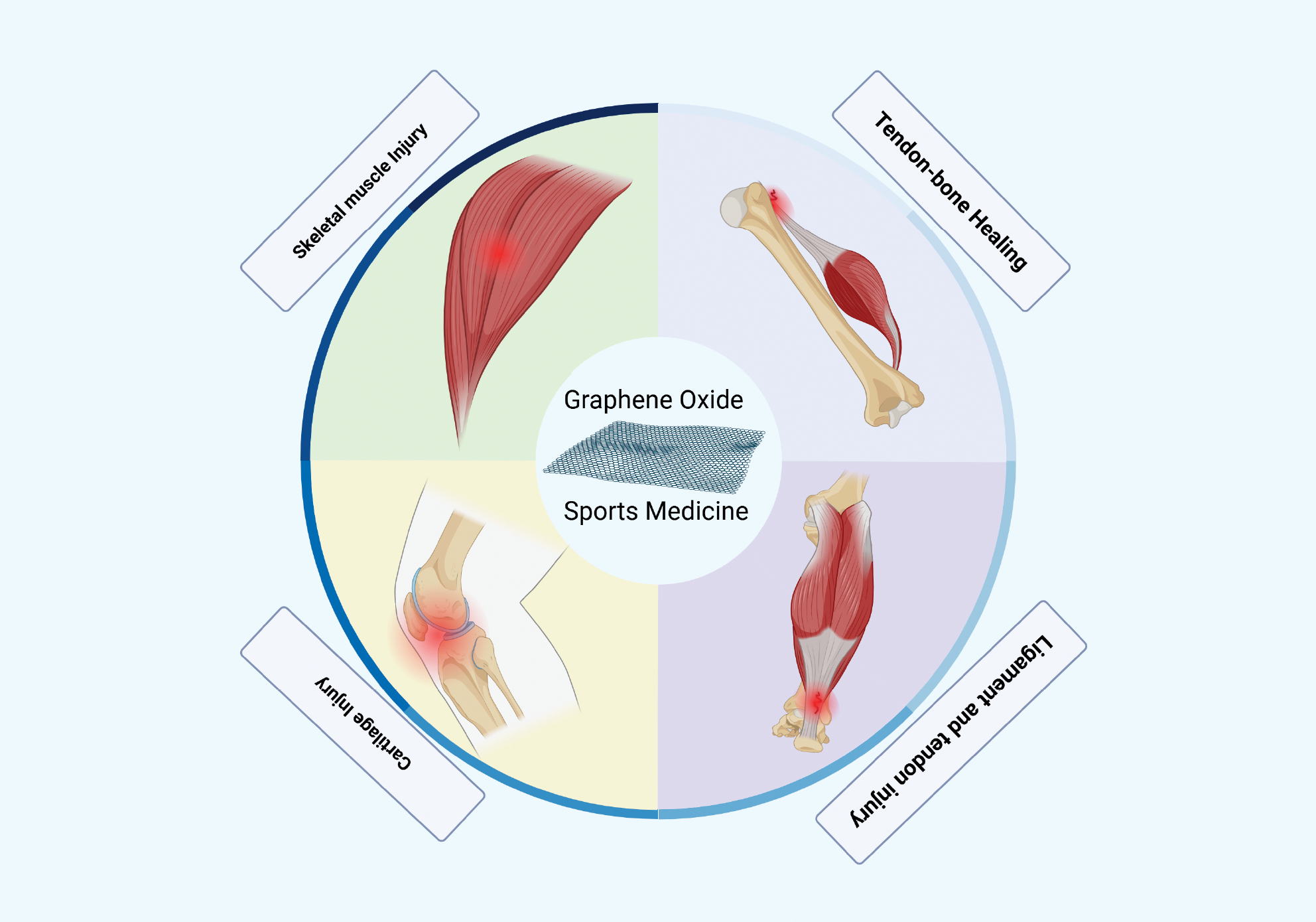
Sports medicine focuses on the prevention and treatment of exercise-related injuries [13, 14], encompassing clinical care, rehabilitation, nutrition, and performance optimization. Sports medicine interrogates physiologic responses to exercise and mechanisms of tissue repair to enhance health and athletic capacity, especially within muscle, bone, joints, ligaments, and tendons (Figure 1) [15–17]. Successful tissue repair in sports medicine typically relies on four pillars: (1) appropriate reparative cells capable of regenerating or replacing damaged tissue; (2) a supportive microenvironment, often provided by a scaffold; (3) relevant biomolecules, such as growth factors that drive proliferation and matrix synthesis; and (4) mechanical cues within the physical microenvironment that guide maturation of the new tissue [18–20]. Biocompatible and tunable biomaterials have therefore drawn substantial interest for the capacity to promote regeneration and wound healing [18–21] and identifying clinically deployable materials has become a central objective [22–24].
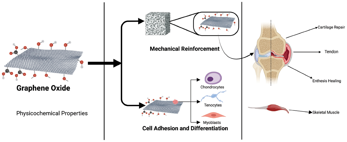
Figure 1 Schematic overview of graphene oxide (GO) applications in sports medicine. The figure illustrates the versatile utility of GO-based biomaterials across various musculoskeletal tissues, including articular cartilage, skeletal muscle, tendon/ligament, and the tendon-bone interface. The central panel highlights the key physicochemical properties of GO, such as mechanical strength, electrical conductivity, and surface functionalization, that enable these tailored therapeutic strategies for tissue regeneration and inflammation modulation.
Compared to traditional biomaterials, such as poly (lactic-co-glycolic acid) [PLGA] or natural polymers (e.g., collagen and alginate), GO-based materials offer a unique combination of advantages [25]. While traditional polymer scaffolds possess good biocompatibility, traditional polymer scaffolds often lack sufficient mechanical strength for load-bearing tissues, like cartilage, and have limited bioactivity [26]. Conversely, metallic or ceramic implants, although robust, lack biodegradability and have a severe modulus mismatch with biological tissues, which potentially causes stress-shielding [27]. The incorporation of GO can significantly enhance the mechanical properties of composite materials at very low concentrations, bringing composite materials closer to the mechanical requirements of native tissues [28]. More importantly, the unique surface chemistry and electrical conductivity of GO provide the material with unprecedented bio-functionality to actively guide cell behavior and tissue regeneration, an attribute often missing in conventional materials [29].
Within this context, GO shows compelling potential for repairing and reconstructing the musculoskeletal system, particularly in cartilage, tendon, ligament, and bone-related injuries. The mechanical strength, electrical conductivity, and thermal stability of GO, coupled with unique biological functions and generally favorable compatibility, make GO an attractive candidate material in sports medicine [1–3]. Nevertheless, opportunities come with challenges. Key issues remain regarding biosafety, precise control of cell–material interactions, and in vivo stability and degradability [6–8]. Thus, despite encouraging prospects, rigorous mechanistic studies and extensive preclinical and clinical testing are still required for translation in sports medicine. Notably, GO serves as a versatile platform for chemical modification, yielding derivatives with properties tailored for specific applications [30]. The most common modification strategy is reduction, which produces rGO. By removing many of the oxygen-containing functional groups, this process partially restores the excellent electrical conductivity of graphene, making rGO particularly promising for the regeneration of electroactive tissues, such as skeletal muscle or nerves [11]. Another key strategy is surface functionalization, in which specific biomolecules (e.g., peptides and growth factors) are attached to the GO surface via covalent or non-covalent bonds [11]. This approach creates materials with enhanced biological specificity, enabling more precise targeting of cells or the controlled release of therapeutic agents to trigger distinct biological pathways. Therefore, this review will not only focus on GO but will also, where appropriate, explore how these key derivatives (rGO and various functionalized GO materials) provide unique solutions to specific challenges in sports medicine. This review synthesizes recent progress on GO-based biomaterials across sports medicine applications to provide guidance and perspective for future research.
Fundamental properties of GO
Chemical structure and physical features
GO possesses a distinctive architecture in which carbon atoms are connected through mixed sp2/sp3 hybridization to form a planar hexagonal network. Abundant oxygen-containing functional groups (primarily hydroxyl, carboxyl, and epoxide moieties) are interspersed on the basal plane and enriched at sheet edges, collectively dictating the reactivity, dispersibility, and interfacial behavior of GO.
Production process
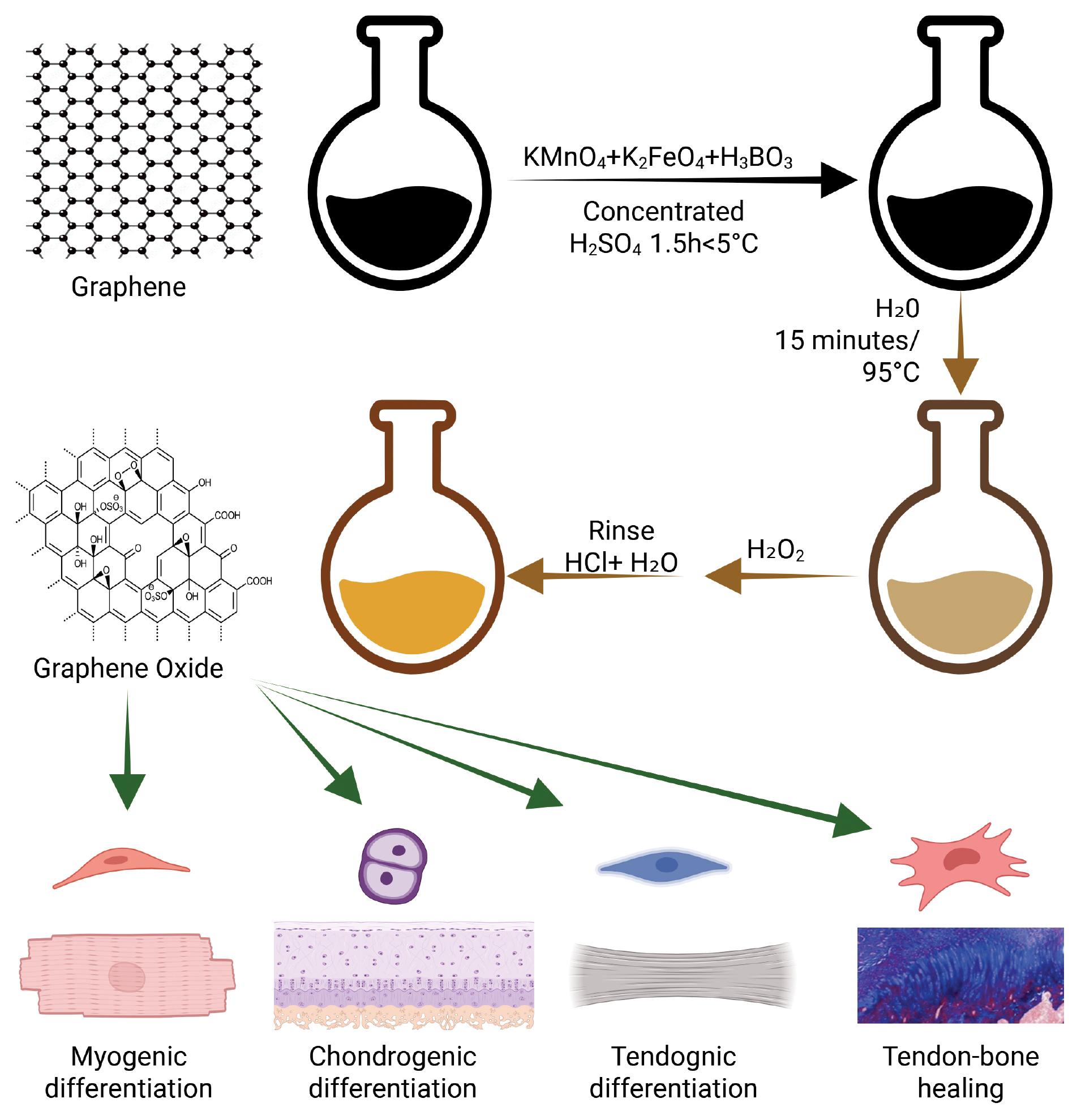
GO is most frequently produced via the Hummers route [31–35]. Briefly, graphite flakes are oxidized under strongly acidic conditions. Graphite is dispersed and cooled to ~5°C in concentrated sulfuric acid, then oxidized using KMnO4 and K2FeO4 for ~1.5 h, followed by further (deep) oxidation with KMnO4. The oxidized material is hydrolyzed with deionized water at 95°C for 15 min, yielding a brown suspension indicative of exfoliated graphite oxide. Residual oxidants and sulfate intermediates are quenched with H2O2 and the product is repeatedly washed with hydrochloric acid and water to remove ionic by-products and obtain purified GO (Figure 2).
Figure 2 From synthesis-to-function: Tailoring graphene oxide (GO) for stem cell differentiation. The schematic depicts the workflow from the synthesis of GO via oxidation of graphite-to-chemical modification (e.g., reduction to rGO and surface functionalization). These physicochemical modifications dictate the interaction of materials with stem cells, thereby directing lineage-specific commitment toward chondrogenic, myogenic, tenogenic, or osteogenic fates through modulation of the cellular microenvironment.
Physical properties
Disruption of the pristine sp2 network imparts GO with pronounced electrical insulation. Depending on the degree of oxidation and reduction state, semiconducting behavior can also emerge. GO exhibits a high specific surface area (up to ~890 m2 g⁻1) together with robust mechanics, which is an effective Young’s modulus on the order of ~1.0 TPa and a fracture strength approaching ~130 GPa [8, 36–38]. These mechanical attributes are critically important for sports medicine applications and enable GO to serve as a highly effective nanofiller that can significantly enhance the mechanical strength and toughness of hydrogels or polymer scaffolds, even at low concentrations, allowing the hydrogels or polymer scaffolds to withstand the physiologic loads experienced by load-bearing tissues, like articular cartilage or tendons [39].
Surface hydrophilicity
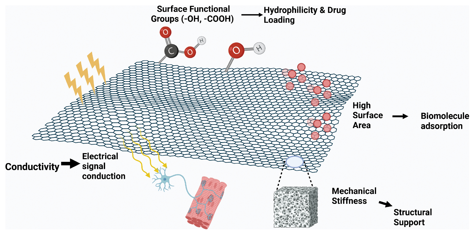
Oxidation converts hydrophobic graphene into hydrophilic GO by introducing hydroxyl, epoxide, and other polar functionalities (primarily carboxyl and carbonyl groups) on the basal plane and sheet edges. These oxygenated groups enable stable dispersion in water and other polar solvents and provide versatile handles for surface chemistry and bioconjugation, facilitating the coupling of proteins, antibodies, and nucleic acids, as well as other bioactive molecules such as growth factors and peptides [6, 8, 40]. This hydrophilicity and abundance of functional groups are particularly advantageous in tissue engineering as the functional groups not only promote the integration of the scaffold material within the aqueous biological environment (Figure 3) but also serve as “anchors” for immobilizing growth factors or adhesion proteins, thereby creating a bioactive microenvironment that can actively guide stem cell differentiation.
Figure 3 Schematic illustration of the key physicochemical properties of graphene oxide (GO) and the corresponding biomedical functions. The diagram maps the intrinsic material attributes of GO nanosheets to specific therapeutic applications in sports medicine: (1) Surface functional groups–the presence of hydroxyl (–OH) and carboxyl (–COOH) groups enhance hydrophilicity and facilitates drug loading. (2) High surface area–provides a vast interface for the efficient adsorption of biomolecules. (3) Mechanical stiffness–enhances the mechanical integrity of composite scaffolds to provide robust structural support. (4) Conductivity–enables electrical signal conduction, which is critical for the regeneration of electroactive tissues such as skeletal muscle and nerves.
Adsorption and interaction mechanisms
GO exhibits strong adsorption toward proteins and antibodies, which can help preserve adsorbates from degradation and is an advantage for protein delivery and biosensing platforms [41, 42]. Protein binding to GO is mediated by multiple non-covalent forces, including hydrophobic interactions, van der Waals forces, electrostatic attraction/repulsion, and hydrogen bonding. Adsorption behavior can shift from Freundlich- to Langmuir-type isotherms as the degree of oxidation increases, reflecting changes in surface heterogeneity and site saturation [43]. The π-electron-rich basal plane also enables π–π stacking with hydrophobic drugs and polymers, supporting the formation of nanocomposites. In addition, covalent conjugation of biomolecules to GO via reactive oxygenated groups can enhance conjugate stability across diverse environments, although care must be taken because covalent coupling can alter protein conformation and function; the use of appropriate crosslinkers can mitigate such effects [7, 44].
The tunable chemistry of GO, large surface area, and high mechanical strength, render GO broadly applicable across biomedicine, including tissue engineering, drug delivery, and biosensing. The ability of GO to form stable aqueous dispersions, present multifunctional binding sites, and undergo controlled chemical modification creates a flexible platform for engineering next-generation bio-interfaces and therapeutic systems.
Biocompatibility
The widespread biomedical use of GO is largely attributable to the favorable biocompatibility. Numerous studies have shown that GO supports the adhesion and proliferation of diverse cell types, including osteoblasts and neurons, making GO an attractive component for tissue-engineered scaffolds [45–49]. Nevertheless, uncertainties remain regarding cytotoxicity and long-term in vivo compatibility and outcomes can vary with production routes and post-processing (e.g., chemical functionalization or physical modification) that modulate surface chemistry and biological responses. Because graphene-based nanomaterials may elicit cytotoxicity and challenge biocompatibility in vivo, GO and GO derivatives must be evaluated in the context of the physicochemical attributes and interactions with the biological milieu.
Cytotoxicity
Biocompatibility is pivotal in determining the in vivo behavior and toxicity profile of nanomaterials. Given the diversity of nano–bio interfaces, intrinsic toxicologic effects are heterogeneous and cannot be generalized. Hydrophobicity and aggregation are key determinants for graphene family materials. Pristine graphene tends to irreversibly aggregate in culture media, causing dose- and time-dependent physical damage to cell membranes and resultant cytotoxicity. By contrast, GO and rGO are more hydrophilic and disperse more readily, which generally lowers cytotoxicity relative to pristine graphene [44, 50]. Although oxygenated surface groups on GO/rGO can promote reactive oxygen species (ROS) generation, improved dispersion markedly reduces aggregation-driven physical injury. Therefore, overall cytotoxicity of GO/rGO is typically lower than pristine graphene once synthetic residues are thoroughly removed. Meticulous purification to eliminate chemical by-products from synthesis is therefore essential to minimize cytotoxicity.
Green synthesis approaches
Greener synthetic strategies have been developed to reduce harmful residuals using biocompatible surfactants and reducing agents to indirectly improve biocompatibility [51]. For example, a trastuzumab-modified, dual-functional graphene nanosheet showed good biocompatibility and supported 3D spheroid growth of human breast cancer cells, illustrating the potential of biofriendly surface chemistry to mitigate toxicity while enabling function [52]. However, in addition to the initial material purity, the long-term biosafety of GO critically depends on degradation and clearance within the body.
Hemocompatibility and immune interactions
Blood–material interactions are critical determinants of in vivo biocompatibility. Pristine graphene, rGO, and GO induce dose-dependent hemolysis with pristine graphene typically showing the strongest hemolytic activity. Pre-incubation with proteins and other surface modifications (such as PEGylation or polymer coating) can substantially diminish this risk [53]. Graphene-based materials may also trigger immune responses, including the release of pro-inflammatory cytokines, such as interleukin-6 (IL-6) and IL-8, and activation of complement pathways that contribute to inflammation and potential chronic sequelae. Notably, macrophages and neutrophils internalize GO more efficiently than hydrophobic graphene, which may influence tissue healing and immune modulation [54]. The interaction between GO and the immune system is a “double-edged sword” [55]. Improper physicochemical properties (e.g., large size and sharp edges) can activate inflammasome pathways, leading to pyroptosis and persistent inflammation [56], while well-designed GO materials can be harnessed for beneficial immunomodulation, as mentioned previously. The net outcome of this interaction is highly dependent on the physicochemical parameters of GO, including lateral size, number of layers, surface charge, and degree of functionalization [57]. For example, smaller, surface-functionalized GO nanosheets tend to exhibit better biocompatibility and lower immunogenicity [58]. Therefore, understanding and precisely controlling the structure-activity relationship between these parameters and the immune response is key to the safe and effective application of GO [55].
In vivo biocompatibility
The biodistribution and fate of graphene nanomaterials are governed by the physicochemical properties and exposure routes. These materials can traverse biological barriers, accumulate in organs, and provoke inflammation or tissue injury [6, 45]. High-dose GO has been shown to cause severe, persistent pulmonary damage in mice, while few-layer graphene accumulates primarily in the lungs after injection, with possible redistribution to the liver and spleen [59]. GO accumulation in organs, such as the spleen, may exert limited histopathologic or functional impact over the long term; some studies report altered T-cell subsets and reduced pro-inflammatory cytokine expression following GO exposure [10]. Designing locally confined, controllable GO-derived materials can restrict systemic accumulation and potential toxicity, thereby broadening applicability, an approach reflected in the growing portfolio of GO-based biomaterials [11, 60, 61].
Recent studies have established specific thresholds for dosage and metabolic kinetics to provide a more quantitative understanding of the biosafety profile. In vitro studies suggested that GO exhibits dose-dependent toxicity with cell viability typically remaining unaffected at concentrations <20 μg/mL, while concentrations >50 μg/mL often induce significant apoptosis [62, 63]. Intravenous administration in mice indicated that a dose of 1–2 mg/kg body weight is generally well-tolerated without acute toxicity, whereas doses >10 mg/kg can lead to pulmonary granuloma formation or organ failure [64]. Furthermore, the pharmacokinetics of GO are strictly governed by the lateral size [65]. Small-sized GO sheets (<50 nm) are rapidly cleared through renal filtration with a blood circulation half-life <1 h, whereas larger sheets are predominantly sequestered by the reticuloendothelial system (RES), primarily in the liver and spleen, where the larger sheets may be retained for > 3 months before gradual clearance [66].
In addition to biodistribution, the GO degradation and clearance mechanisms are critical to long-term safety. Studies have shown that GO is not entirely non-biodegradable. GO can be degraded by specific enzymes in a biological environment [67]. For example, myeloperoxidase (MPO), an enzyme secreted by immune cells, like neutrophils, can effectively catalyze the degradation of GO in the presence of hydrogen peroxide (H2O2) [68]. This process breaks down GO into smaller, soluble fragments that can be cleared from the body, potentially via the renal system [69]. The rate of degradation is influenced by the size, degree of oxidation, and surface chemistry of the GO sheets [69–71]. Therefore, a rational design of the physicochemical parameters of GO holds promise for achieving controllable degradation behavior, thereby minimizing long-term accumulation and potential risks in vivo.
Bioactivity
Chemical functionalization is a powerful lever for enhancing both biocompatibility and bioactivity. Peptides and other biomolecules grafted onto GO can strengthen cell–material interactions and direct specific cell behaviors, including lineage commitment [72, 73]. In addition, GO’s large surface area and amenability to modification make it a versatile carrier in drug-delivery systems, capable of loading and controlled release of a broad range of bioactive agents [9].
Collectively, thanks to its unique chemistry, tunable surface, and generally favorable biocompatibility, GO is a multifunctional platform with substantial promise in biomedicine. Nevertheless, successful translation requires deeper interrogation of potential challenges, especially cytotoxicity drivers, immune interactions, biodistribution, and long-term stability, and the development of standardized, application-specific purification and functionalization workflows to ensure safety and efficacy in vivo.
Mechanisms of GO-mediated cellular modulation
The regenerative efficacy of GO-based biomaterials extends beyond the role as passive structural scaffolds. The regenerative efficacy of GO-based biomaterials is fundamentally driven by dynamic physicochemical interactions at the material-cell interface [74]. Understanding these mechanisms is crucial for elucidating how GO directs cell fate, as follows: 1) surface chemistry and the protein corona – The high surface energy and abundant oxygen-containing functional groups (e.g., hydroxyl and carboxyl groups) on GO nanosheets facilitate the rapid adsorption of proteins from the biological environment [75]. This formation of a “protein corona,” which is often enriched with adhesive proteins like, fibronectin, serves as the primary biological interface [75]. The primary biological interface bridges the inorganic material and the cell, directly modulating integrin binding and subsequent focal adhesion formation. 2) mechanotransduction and topography – Cells are highly sensitive to the physical cues of the microenvironment [76]. The unique nanotopography of GO is characterized by ridges, ripples, and edges. When combined with tunable stiffness, the GO nanotopography exerts physical forces on the cytoskeleton [77]. These mechanical stimuli are transduced intracellularly, notably triggering the nuclear translocation of mechanosensors, such as YAP/TAZ [78]. This process translates physical cues into biochemical signals that regulate gene expression related to proliferation and differentiation [79]. 3) ROS scavenging and microenvironment regulation – While high concentrations of GO can induce oxidative stress, controlled amounts of GO and GO derivatives (especially rGO) exhibit enzyme-mimetic properties [80]. This capability allows for the scavenging of excessive ROS, thereby protecting cells from oxidative damage and creating a permissive microenvironment for tissue regeneration.
Collectively, these surface, mechanical, and chemical interactions converge to activate canonical signaling pathways, such as Wnt/β-catenin, TGF-β/Smad, and MAPK/ERK, which ultimately dictate the lineage commitment of stem cells and the functional restoration of tissues [81–83].
Applications of GO in sports medicine
Overview: multifunctional applications of GO in musculoskeletal tissue engineering
The unique physicochemical properties of GO, including exceptional mechanical strength, vast specific surface area, tunable electrical conductivity, and abundant surface chemistry, render GO a highly promising and versatile platform for addressing key challenges in sports medicine and orthopedic tissue engineering [84]. This chapter will explore the specific applications of GO across a range of musculoskeletal regeneration contexts, beginning by examining the role of GO in cartilage repair, in which GP is primarily leveraged as a mechanical reinforcing agent and a carrier for bioactive molecules to create functional scaffolds (Section 3.2) [26]. Subsequently, the use of GO in skeletal muscle regeneration will be investigated, focusing on how electrical conductivity and nanotopography are critical for guiding the formation and maturation of electroactive muscle tissue (Section 3.3) [85]. The discussion will then extend to ligament and tendon repair, highlighting how GO can enhance the toughness and load-bearing capacity of engineered constructs for high-strain environments (Section 3.4) [86]. In addition to the structural roles of GO, the capacity of GO as a bioactive modulator in regulating post-traumatic inflammation will be explored, in which antioxidant and immunomodulatory properties can steer the injury microenvironment toward a pro-reparative state (Section 3.5) [87]. Finally, the complex challenge of tendon–bone healing will be addressed, illustrating how GO-based strategies facilitate the recreation of a functional enthesis by providing mechanical stability, directing cell differentiation, and controlling growth factor release (Table 1) (Section 3.6) [88]. Collectively, these applications demonstrate that GO is evolving from a passive scaffold material into an active, multifunctional component in advanced regenerative strategies for sports-related injuries.
Table 1 Summary of Graphene Oxide (GO) Applications in Tendon–bone Healing
| Study / Author | Material System / Scaffold Design | Experimental Model | Key Findings & Outcomes |
|---|---|---|---|
| Su et al. [140] | GO-doped PLGA nanofiber membranes | Rabbit model |
|
| Yoon et al. [60] | GO–alginate sheet scaffold | Rat rotator cuff tendon–bone interface (TBI) |
|
| Bao et al. [143] | GO combined with platelet-rich plasma (PRP) gel | Rabbit model |
|
GO in cartilage injury repair
Articular cartilage has negligible intrinsic regenerative capacity, making durable repair challenging and complicating management of cartilage-related disorders, such as osteoarthritis [89–92]. Tissue engineering has therefore emerged as a promising route. Incorporating graphene-based materials into cartilage scaffolds can markedly enhance electrical conductivity and mechanical robustness, leveraging the electro-mechanical attributes of graphene [93–97].
To achieve effective chondral regeneration, investigators have explored GO and GO-containing bio-composites to direct mesenchymal stem cells (MSCs) toward chondrogenesis with encouraging results [98–100]. GO augments chondrogenic differentiation efficiency and higher scaffold porosity, which increases GO loading, can further accelerate lineage commitment [98–100]. GO is frequently combined with biocompatible polymers (e.g., polyvinyl alcohol and chitosan) in cartilage repair to form nanocomposite hydrogels that mimic cartilage extracellular matrix (ECM). These composites exhibit cytocompatibility, responsiveness to external stimuli, and favorable biomechanics. GO incorporation significantly strengthens the matrix and improves durability, which is critical for load-bearing cartilage (Table 2) [95]. Moreover, the surface functional groups of GO interact with bioactive molecules (e.g., proteins and growth factors), promoting stem-cell adhesion and growth [47]. Such composites support cell proliferation and stimulate production of cartilage-specific macromolecules, including collagens and proteoglycans, which are essential for hyaline cartilage formation.
Table 2 Summary of Graphene Oxide (GO) Applications in Cartilage Injury Repair
| Study (Ref.) | Material Composition & Design | Experimental Model | Key Mechanisms & Therapeutic Outcomes |
|---|---|---|---|
| Trucco et al. [94] | Bilayer hydrogel: GO-doped gellan gum + poly(ethylene glycol) diacrylate (PEGDA) | In vitro (cartilage mimetic) |
|
| Ogene et al. [104] | Nanomaterial: GO nanosheets (interaction study) | In vitro (human chondrocyte line) |
|
| Liu et al. [49] | Injectable: Umbilical cord MSCs (UCMSCs) + GO-particle lubricant | In vivo (rabbit papain-induced OA model) |
|
| Wang et al. [48] | Injectable: UCMSCs + GO-particle lubricant | In vivo (modified Hulth OA model) |
|
| Cheng et al. [96] | Scaffold: 3D printed GO-containing scaffolds | In vitro & In vivo |
|
| Li et al. [105] | Meniscal scaffold: GO coated with tannic acid, Sr2⁺, and silk fibroin | In vivo (meniscal/osteochondral) |
|
| Jiao et al. [106] | Drug delivery: Gelatin-reduced GO (rGO@Ge) delivering Kartogenin (KGN) | In vitro (rat ADSCs) |
|
| Abedin Dargoush et al. [107] | Bilayer construct: PEGDA (top) + GelMA (bottom) with nHA and GO | In vitro & In vivo |
|
Mechanistically, GO nanosheets can adsorb transforming growth factor (TGF)-β and fibronectin, while preserving the bioactive conformations, which are key to driving chondrogenesis [47]. In addition to single-component systems, hybrid nanocomposites pair GO with other nanoparticles to boost mechanics. For example, GO–chitosan scaffolds combine GO reinforcement with chitosan biocompatibility and have been shown to stimulate proliferation and improve morphology of human articular chondrocytes [101, 102]. Additional GO-based combinations, such as with polyvinyl alcohol, PEGMA-b-PCL block co-polymers, or chondroitin sulfate, have yielded scaffolds with favorable cell morphology, cytocompatibility, and in vivo repair of cartilage defects [103].
Trucco et al. engineered GO-doped gellan gum–poly(ethylene glycol) diacrylate (PEGDA) bilayer hydrogels to emulate the mechanical and lubricative functions of articular cartilage [94]. GO-containing hydrogels display excellent compressive and shear properties and lubrication comparable to native cartilage, are non-cytotoxic in vitro, and support cell proliferation and chondrocytic differentiation, which highlights the promise for joint restoration. Ogene et al. reported that GO increases plasma membrane tension and activates the TGF-β pathway in a human chondrocyte line, enhancing SMAD2/3 phosphorylation and downstream gene expression [104]. Fluorescence lifetime imaging suggested strong GO–membrane interactions that trigger mechanoresponsive signaling and endogenous TGF-β activation.
Liu et al. evaluated umbilical cord MSCs (UCMSCs) delivered with a GO-particle lubricant in a rabbit papain-induced osteoarthritis model [49]. The combination reduced inflammatory cytokines in synovial fluid and serum, ameliorated subchondral osteopenia, and promoted cartilage repair, indicating that GO particles can provide superior lubrication while acting as a stem-cell carrier to enhance therapeutic efficacy. Similarly, Wang et al. showed that UCMSCs plus a GO-particle lubricant markedly decreased nitric oxide (NO), interleukin (IL)-6, and tumor necrosis factor (TNF)-α in a modified Hulth model, while increasing glycosaminoglycan (GAG) and type II collagen (COL-II), thereby improving the joint biochemical milieu and facilitating cartilage repair [48]. GO particles alone had a limited effect and UCMSCs alone provided partial benefit.
Additive manufacturing has also been leveraged. Cheng et al. used 3D printing to fabricate GO-containing scaffolds that enhanced chondrocyte proliferation and collagen synthesis with favorable biocompatibility and mechanics in vitro and in vivo, although optimization of GO content and mechanistic dissection remain needed [96]. Li et al. developed a GO-based meniscal scaffold coated with tannic acid, Sr2⁺, and silk fibroin, which demonstrated anti-inflammatory and antioxidant (anti-ROS) effects that protect cartilage and delay osteoarthritis progression in vivo with good safety [105]. Jiao et al. employed gelatin-reduced GO nanosheets (rGO@Ge) to deliver kartogenin (KGN), promoting chondrogenic differentiation of rat adipose-derived stem cells with concomitant modulation of autophagy [106]. Abedin Dargoush et al. proposed a functional bilayer osteochondral construct (PEGDA top layer and GelMA bottom layer) incorporating nano-hydroxyapatite (nHA) and GO [107]. The scaffold improved cell adhesion, proliferation, and lineage-specific differentiation in vitro and in vivo, while providing suitable biocompatibility and mechanics for coordinated cartilage and bone regeneration.
Collectively, these studies indicated that GO can achieve the following: (i) reinforce scaffold mechanics and lubrication; (ii) present and/or potentiate chondrogenic cues (e.g., via TGF-β adsorption and mechanoactivation); (iii) serve as a carrier/lubricant to synergize with stem-cell therapies; and (iv) integrate with advanced fabrication to engineer ECM-mimetic architectures. Remaining priorities include defining optimal GO loading/functionalization, long-term in vivo safety, and standardized endpoints for translating GO-based cartilage therapies.
Applications of GO in skeletal muscle regeneration
Skeletal muscle is a contractile tissue composed of highly aligned bundles of myofibers and has a central role in locomotion and postural support [108, 109]. Severe functional impairment can arise from trauma, myopathies, or tumor ablation [19, 110–113]. To repair such injuries, investigators are actively exploring stem cell–nanomaterial strategies in which scaffolds guide cell fate and enhance differentiation capacity, thereby promoting skeletal muscle regeneration [73, 114, 115]. Owing to the favorable physical properties (moderate electrical conductivity [~0.6 S/m], ultralow density, flexibility, and high mechanical strength) graphene-derived materials are considered promising myogenic substrates. Specifically, GO-based constructs have repeatedly been shown to support myogenic precursor adhesion, proliferation, and differentiation (Table 3) [116, 117].
Table 3 Summary of Graphene Oxide (GO) Applications in Skeletal Muscle Regeneration
| Study / Author | Material System / Scaffold Design | Experimental Model / Context | Key Findings & Mechanisms |
|---|---|---|---|
| Shin et al. [118] | Mixed matrices of GO with PLGA and collagen | C2C12 myoblasts |
|
| Ku and Park [119] | GO and rGO immobilized on amine-functionalized glass | C2C12 myoblasts |
|
| Jo et al. [120] | GO/polyacrylamide (GO/PAAm) composite hydrogels | C2C12 myoblasts |
|
| Chaudhuri et al. [45] | Dielectric and semiconducting thin GO films | Cord blood–derived human MSCs (CB-hMSCs) |
|
| Aparicio-Collado et al. [117] | Semi-interpenetrating nanohybrid hydrogel (alginate + PCL + rGO) | In vitro (electroactive tissue focus) |
|
| Keremidarska-Markova et al. [121] | GO and PEG-modified GO (GO-PEG) + NIR irradiation | Ex vivo (frog heart), C2C12, Rat liver |
|
| Kang et al. [122] | 3D-printable hydrogel (Gelatin-GHPA + GO) | C2C12 myoblasts |
|
| Ko et al. [116] | rGO sheets in a valveless biohybrid pump | Engineered skeletal muscle (bio-actuation) |
|
| Zhang et al. [123] | Gelatin/rGO conductive micro-cryogel (injectable) | Mouse tibialis anterior injury model |
|
| Choi et al. [73] | Nanoscale GO (sGO) coatings on nanopillar arrays | In vitro (micro/nanotopography) |
|
Shin et al. demonstrated that mixed matrices of GO with PLGA and collagen significantly enhanced proliferation and adhesion of C2C12 myoblasts and further promoted myogenic differentiation, evidenced by multinucleated myotube formation and increased myosin heavy chain (MHC) expression [118]. Consistently, Ku and Park reported that GO- and rGO-modified glass substrates modulate C2C12 differentiation [119]. GO immobilized on amine-functionalized glass with rGO produced by hydrazine reduction markedly improved cell adhesion, proliferation, and myogenesis, which was attributed to serum-protein adsorption and nanoscale topography. GO increased multinucleated myotube formation, upregulated myogenic regulators (e.g., MyoD), and boosted myogenic protein synthesis relative to unmodified and rGO substrates. The wrinkled GO surface likely anchors proteins and cells more effectively. Moreover, the higher density of GO oxygenated groups (carboxyl, hydroxyl, and epoxide) enhances serum-protein adsorption, and in turn, myogenic differentiation, which underscores the promise of GO in muscle tissue engineering.
Jo et al. showed that GO/polyacrylamide (GO/PAAm) composite hydrogels promote C2C12 growth, adhesion, and differentiation [120]. Further reduction of the composite enhanced cell–substrate interactions, likely by improving electrical conduction and simplifying intercellular electrical communication that cues myogenesis. Direct differentiation of cord blood–derived human mesenchymal stem cells (CB-hMSCs) into human skeletal muscle cells (hSkMCs) on dielectric and semiconducting thin GO films yielded superior viability, aspect ratio, and expression of myogenic markers compared to control surfaces, highlighting the translational potential of GO for muscle regeneration [45].
Aparicio-Collado et al. developed a semi-interpenetrating nanohybrid hydrogel based on sodium alginate (SA), polycaprolactone (PCL), and rGO nanosheets to engineer conductive, bioactive 3D matrices [117]. rGO markedly increased conductivity, which is critical for excitable tissues, while the network architecture improved physical integrity and biofunction, including adhesion and myogenic differentiation. This finding suggested utility for regenerating electroactive tissues, such as skeletal muscle. Keremidarska-Markova et al. examined GO and PEG-modified GO (GO-PEG) under near-infrared (NIR) irradiation in a multi-scale safety/efficacy appraisal [121]. GO/GO-PEG altered rhythmic contractions in ex vivo frog hearts and elevated ROS in C2C12 cells (attenuated after NIR). GO/GO-PEG increased diamine oxidase activity (potentiated by NIR) in rat liver. While intact mitochondrial function was unaffected, GO reduced ATPase activity in freeze–thawed mitochondria in a concentration-dependent manner. These data illustrated the complex bioeffects of GO derivatives and informed safety assessment for biomedical use.
Printable, GO-containing bioinks are also emerging. Kang et al. reported a 3D-printable hydrogel composed of gelatin–hydroxyphenylacetic acid (GHPA) and GO, crosslinked in situ via a dual-enzyme reaction, which created a supportive microenvironment that drove C2C12 differentiation into myocytes and myotubes, emphasizing the potential of GO-derived bioinks for muscle tissue engineering and regeneration [122]. From a bioactuation perspective, Ko et al. used rGO sheets to enhance the performance of a valveless biohybrid pump driven by engineered skeletal muscle [116]. rGO anchoring delayed early downregulation of connexin-43 (a barrier to myogenesis) and RNA-seq revealed upregulation of positive myogenic regulators (e.g., troponins) with suppression of negative regulators. rGO-modified myotubes generated ~3-fold higher contractile force, yielding substantially increased flow speed and rate, advancing physiologically relevant engineered muscle and high-power bio-machines.
Zhang et al. created a gelatin/rGO conductive micro-cryogel at the injectable-matrix scale that was designed as a myogenic cell carrier [123]. The rGO-mediated conductivity promoted myoblast proliferation and differentiation and in a mouse tibialis anterior injury model the micro-cryogels effectively delivered myogenic cells, which significantly improved muscle repair and regeneration. Choi et al. reported that nanoscale GO (sGO) coatings on nanopillar arrays in micro/nanotopographic systems enhanced adhesion and spreading of myoblasts and efficiently guided myogenic differentiation into mature myotubes, providing a rapid, effective in vitro platform for generating muscle cells [73].
In brief, across 2D films, hydrogels, printable bioinks, and injectable microgels, graphene-based (especially GO-based) materials provide electroconductive, mechanically robust, and protein-interactive interfaces that (i) strengthen myogenic precursor adhesion and proliferation, (ii) potentiate lineage commitment and myotube maturation, and (iii) enable functional bioactuation. Priorities for translation include defining optimal GO/rGO loading and oxidation state, standardizing electrical/mechanical cues, and thoroughly evaluating ROS-related effects and long-term biosafety in clinically relevant models.
Applications of GO in ligament and tendon repair
Injuries to ligaments and tendons remain a major challenge in sports medicine and clinical practice. These tissues possess limited intrinsic regenerative capacity. Therefore, conventional approaches, such as suture repair or grafting, often fail to restore native mechanics and function fully [124–126]. Post-injury scarring and fibrosis can further compromise functional recovery and elevate the risk of re-injury [125, 127, 128]. Consequently, current research focuses on advanced biomaterials and tissue-engineering strategies to more effectively drive ligament and tendon regeneration (Table 4).
Table 4 Summary of Graphene Oxide (GO) Applications in Ligament and Tendon Repair
| Study / Author | Material System / Scaffold Design | Experimental Model / Context | Key Findings & Mechanisms |
|---|---|---|---|
| Sarkar et al. [129] | Poly(acrylic acid) hydrogel physically and chemically crosslinked by 2D GO | High-strain tissue engineering (e.g., ligaments and myocardium) |
|
| Saveh Shemshaki et al. [132] | Graphene nanosheets (GnPs) dispersed in poly-L-lactic acid (PLLA) | Massive rotator cuff tear (MRCT) / rat model & in vitro |
|
| Barzegar et al. [133] | Hybrid hydrogel: Gelatin integrated with hyperbranched polyglycerol (HPG) grafted onto rGO and MoS2 | Achilles tendon repair |
|
Sarkar et al. described a poly(acrylic acid) hydrogel physically and chemically crosslinked by 2D GO that exhibited exceptional toughness and extensibility without sacrificing modulus [129]. GO, as a multifunctional crosslinker, strengthens interchain bonding, yielding hydrogel mechanics suitable for constructing artificial tissues that experience high strains, such as ligaments and myocardium, highlighting the promise for load-bearing soft-tissue repair.
Massive rotator cuff tears (MRCTs) present a formidable clinical challenge. MRCTs are typified not only by the tendon rupture but also by severe secondary muscle degeneration, including atrophy, fatty infiltration, and fibrosis [130]. These pathologic changes in the muscle are difficult to reverse with standard surgery, which primarily offers a mechanical fix, leading to high retear rates and unsatisfactory functional outcomes [131]. To address degenerative changes associated with MRCTs, Saveh Shemshaki et al. developed an electrically conductive matrix composed of graphene nanosheets (GnPs) dispersed in poly-L-lactic acid (PLLA) [132]. MRCTs are difficult to reverse with standard surgery. The GnP matrix elevates intracellular Ca2⁺ in myocytes, promotes myotube formation, and suppresses adipogenesis in vitro and reduces fatty infiltration and fibrosis, attenuates muscle atrophy, and improves tendon morphology and tensile properties in a rat MRCT model (Figure 4).
Figure 4 In vivo therapeutic evaluation of graphene nanoplatelet (GnP) matrices in a chronic massive rotator cuff tear model. Histologic cross-sections (H&E staining) of the infraspinatus muscle at 32 weeks post-injury demonstrate the regenerative capacity of the scaffold. The No Repair (NR-32) and Suture Repair (S-32) groups exhibited severe fatty infiltration (white spaces) and muscle atrophy. In contrast, the GnP Matrix (G-32) group effectively preserved muscle architecture and minimized fatty degeneration, resembling Native (N-32) tissue. Figure 4 was reproduced from reference 89 with permission from the author (s). Copyright 2022, Reference: Saveh Shemshaki, N. et al., 2022 [132].
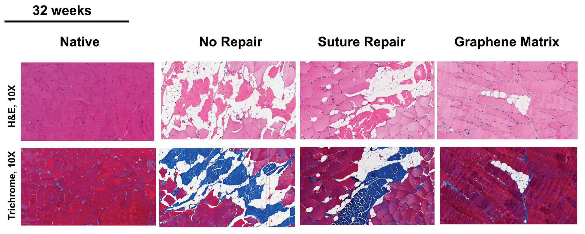
Long-term implantation revealed favorable biocompatibility with no evident visceral toxicity, suggesting translational potential for mitigating MRCT-related degeneration, lowering retear rates, and improving surgical outcomes. Therefore, this approach represents a significant paradigm shift, moving beyond simple mechanical repair to actively modulate the muscle biological environment, thereby targeting the root cause of surgical failure in MRCT treatment.
Barzegar et al. created a multifunctional hybrid hydrogel for Achilles tendon repair by chemically grafting hyperbranched polyglycerol (HPG) onto rGO and MoS2, then integrating the hybrid with a gelatin matrix [133]. The scaffold significantly enhanced tendon regeneration, reduced inflammation, and imparted antibacterial activity in vivo, collectively supporting utility for tendon tissue engineering.
Applications of GO in modulating post-traumatic inflammation
Sports injuries typically trigger an intense inflammatory response [134]. A central therapeutic challenge is to guide the immune microenvironment from an early pro-inflammatory state (dominated by M1 macrophages) toward a pro-reparative state (with resolving inflammation), thereby preventing chronic inflammation and tissue fibrosis.
As outlined in Chapter 2, GO-based biomaterials can act as modulators of this process, although the effects are highly dependent on physicochemical parameters and context. Leveraging the intrinsic antioxidant capacity (see Section 2.2.3), GO-containing constructs can dampen the early burst of ROS and mitigate oxidative stress–related damage. In parallel, tuning GO size, oxidation state, and surface chemistry has been shown to attenuate excessive pro-inflammatory activation and improve hemocompatibility [135]. Across studies, more consistent observations include reduced pro-inflammatory cytokine output and the establishment of a microenvironment that is more permissive to tissue repair [136].
Accordingly, the application of GO in sports medicine extends beyond passive structural roles. By tempering excessive inflammation and oxidative stress in the post-traumatic environment, GO-based systems offer a promising strategy to improve the quality and efficiency of recovery from sports injuries.
Applications of GO in tendon–bone healing
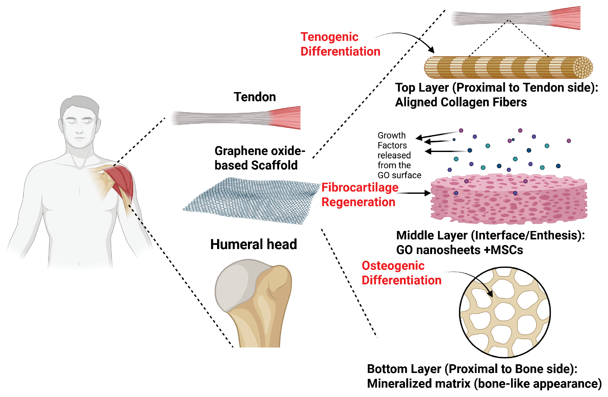
Tendon–bone healing, the re-establishment of a functional enthesis after injury, is pivotal in sports trauma and orthopedic reconstruction. However, the process is frequently hindered by slow repair, fibrotic interfaces, osteopenia, and incomplete recovery of biomechanical properties [137–139], which contribute to suboptimal function and elevated re-injury risk [140–142]. While suture and graft techniques are standard, both often fall short of regenerating a native-like enthesis, motivating the development of GO-enabled materials and strategies (Figure 5).
Figure 5 Schematic illustration of graphene oxide (GO)-based biomaterials for tendon-to-bone interface regeneration. The schematic demonstrates the anatomic placement and the biological mechanism of the GO scaffold in rotator cuff repair. (Left & middle) The GO-based scaffold is implanted at the enthesis defect site, bridging the tendon and the humeral head. (Right) The scaffold facilitates a tri-phasic regeneration process: Top layer (Tendon side): Promotes tenogenic differentiation and guides the formation of aligned collagen fibers. Middle layer (Interface): Incorporates GO nanosheets and MSCs to release growth factors, fostering fibrocartilage regeneration to recreate the natural enthesis transition. Bottom layer (Bone side): Induces osteogenic differentiation and matrix mineralization to ensure firm osseointegration.
Su et al. investigated GO-doped PLGA nanofiber membranes in a rabbit model and reported that intraoperative application of GO–PLGA significantly enhances cell proliferation and osteogenic differentiation, strengthens tendon–bone integration, and increases biomechanical properties compared to PLGA alone [140].
In a 2022 study, Yoon et al. demonstrated that a GO–alginate sheet scaffold markedly improved healing at the rat rotator cuff tendon–bone interface (TBI) [60]. GO contributed mechanical reinforcement, supporting cell activities critical for repair, while alginate provided a cyto-compatible milieu, together preserving TBI structural integrity and fostering cell–cell interactions.
Complementing these findings, Bao et al. showed that combining GO with platelet-rich plasma (PRP) optimized the PRP gel microarchitecture and mechanics and stabilized growth-factor release, thereby improving tendon–bone healing and biomechanics in a rabbit model relative to other treatments [143].
Collectively, these studies underscored the potential of GO-based composites, all of which are crucial for durable joint function, as follows: (i) enhance the local microenvironment; (ii) reinforce mechanical coupling across the enthesis; and (iii) promote biological integration. Future work should deepen mechanistic insight (ideally via multi-omics) to clarify how GO modulates cell fate decisions and immune cues during enthesis regeneration and to define dose, oxidation state, and functionalization parameters for safe, effective clinical translation.
Conclusions and future perspectives
As a two-dimensional material with distinctive physicochemical properties, GO exhibits wide-ranging promise for biomedical applications [6, 45, 116, 144, 145]. This review summarized GO chemistry and materials attributes with emerging uses in sports medicine and outlined priorities for future investigation.
GO has diverse uses due to generally favorable biocompatibility and tunable surface chemistry. Notable progress has been made in cartilage repair, skeletal muscle regeneration, ligament/tendon repair, and tendon–bone healing [94, 123, 133, 143]. GO strengthens hydrogel matrices in cartilage engineering, thereby enhancing cell adhesion and growth and improving the local microenvironment for chondrocytes [93–96]. For example, composites of GO with biocompatible polymers, such as collagen, have supported robust chondrocyte proliferation and differentiation, an important step for hyaline cartilage regeneration. GO electrical conductivity in skeletal muscle can emulate the electrophysiologic context of muscle contraction, promoting myogenic maturation and functional integration [42, 73, 117, 121]. At the same time GO nanoscale architecture offers a supportive 3D framework that facilitates nascent myofiber formation and alignment. GO-reinforced nanofibrous scaffolds in ligament and tendon repair mimics the native extracellular matrix and improves cell attachment and migration, which are key to accelerating repair, while the enhanced mechanics help the construct withstand physiologic loads and maintain structural stability during healing [129, 133]. GO has been used to promote biochemical and mechanical coupling across the interface within tendon–bone (enthesis) repair, improving TBI quality and biomechanics [60, 140, 143]. The surface functional groups can sequester growth factors, guide lineage commitment, and support formation of a graded mineralized fibrocartilage.
Despite these advances, several issues must be resolved to accelerate translation. First, biocompatibility and long-term in vivo stability require more thorough evaluation, including the risks of chronic and/or inflammatory responses following implantation [146, 147]. Current understanding of late-phase host responses to GO are incomplete, calling for standardized long-term safety studies across doses, oxidation states, flake sizes, and routes of administration [6, 102, 58]. Second, although GO provides superior mechanics and bioactivity relative to many matrices, precisely controlling the biochemical cues to meet the divergent requirements of different tissues is challenging [47, 105, 107]. Third, there is a need to engineer next-generation GO-derived biomaterials that more faithfully recapitulate the human biomechanical environment, e.g., via advanced bioprinting strategies to build constructs with complex architectures and spatially programmed functions [148, 149]. Finally, mechanistic insight is limited. Future work should integrate multi-omics (genomics, proteomics, and single-cell and spatial profiling) to interrogate how GO shapes cellular fate decisions, immune modulation, and matrix remodeling across scales [150–152].
In addition to general biosafety, the most pressing challenge lies in the inherent heterogeneity of GO surface chemistry [153]. The co-existence of hydroxyl, epoxy, and carboxyl groups in varying ratios creates a chemically anisotropic surface where different functional domains interact distinctly with proteins, cells, and tissues [153]. This heterogeneity becomes even more problematic when GO undergoes spontaneous reduction in biological environments [154]. The presence of ascorbic acid, glutathione, or enzymatic systems can progressively alter the hydrophilicity, conductivity, and bioactivity over the therapeutic timeframe [155]. Mechanical stress and inflammatory mediators make these chemical transformations particularly unpredictable in sports medicine applications, which complicates efforts to maintain consistent therapeutic effects [156].
Current GO production methods face significant limitations in achieving the precision needed for clinical use [157]. While Hummers’ method and modifications remain the industrial standard, GO with broad distributions in lateral size and C/O ratios are invariably produced [158]. These variations from batch-to-batch directly impact biological responses. Surface modification strategies often fail to achieve uniform coverage because functional groups are irregularly distributed across the GO surface [159]. This feature results in exposed patches that can trigger unwanted inflammatory responses. Another fundamental problem is that mechanical and chemical properties cannot be tuned independently. Increasing oxidation improves dispersibility but weakens the material mechanically, while reduction enhances conductivity at the cost of colloidal stability [160].
Future breakthroughs will require rethinking how GO is synthesized and engineered at multiple scales. Electrochemical and photochemical oxidation methods offer better control over size and oxidation distributions compared to traditional chemical approaches. Real-time monitoring of C/O ratios during synthesis could enable unprecedented quality control [161]. One promising direction involves developing GO derivatives with stable oxidation states that resist biological reduction [153]. For sports medicine specifically, the field needs materials that respond predictably to mechanical loading or inflammatory signals [74]. GO composites that change conductivity or release drugs in response to physiologic stress could revolutionize adaptive therapeutics. Most importantly, we must move beyond empirical optimization to establish clear relationships between GO structural parameters and the biological effects. Only then can we design materials rationally for specific therapeutic applications.
Despite promising preclinical findings, translating these results to the clinic is a formidable challenge. Most current data rely on small animals, like rodents, which simply cannot mimic the extreme load-bearing environment and complex biomechanics of the human musculoskeletal system. In addition to biomechanics, the long-term biological fate of these nanomaterials is a major blind spot. While short-term biocompatibility is often reported, data on how these materials behave over years of residence in the body are lacking. Questions remain about whether the materials degrade completely, accumulate in vital organs, or trigger chronic, low-grade inflammation that could compromise tissue healing. Regulatory bodies are unlikely to approve such therapies without rigorous evidence ruling out delayed toxicity. Future success depends on shifting focus from short-term efficacy to long-term safety monitoring in large-animal models. In summary, GO is a multifunctional platform with substantial potential across sports medicine. Priorities for the field include the following: (i) optimizing biocompatibility and long-term safety; (ii) exerting tighter control over surface chemistry and biochemical signaling; and (iii) developing structurally sophisticated, GO-based composites tailored to the mechanical and biological demands of specific tissues. Addressing these points will help realize the clinical utility of GO and broaden the impact in tissue engineering, regenerative medicine, and disease therapy.
Data availability statement
All data are available in the main text or the supplementary materials. The materials generated are available from the lead contact upon reasonable request.
Ethics Statement
No direct interactions with human or animal subjects were involved. Therefore, ethical approval and informed consent were not required.
Author contributions
Equal contribution. Mengran Xi (MX), Renwen Wan (RW), and Wei Luo (WL) contributed equally to this work.
- Conceptualization: Zhiwen Luo (ZL), Shiyi Chen (SC), Jiajun Qiu (JQ).
- Project administration: ZL, SC.
- Supervision: ZL, SC, JQ.
- Methodology (scope definition, search strategy, inclusion/exclusion criteria): MX, RW, WL, with guidance from ZL and JQ.
- Investigation / Literature search and curation: MX, RW, WL, Yanwei He (YH), Xinting Feng (XF), Tianqi Wang (TW).
- Data curation (reference management, figure/table sourcing, citation checking): MX, RW, WL, YH.
- Writing – original draft: MX, RW, WL (primary); YH, XF, TW (section drafts and figure captions).
- Writing – review & editing (scholarly revision, clinical framing, and materials science accuracy): SC (clinical and sports-medicine perspectives), JQ (materials science and nanomaterials), ZL (overall synthesis and positioning).
- Visualization (schematics and graphical summary): WL, XF, TW, with input from JQ.
- Validation (cross-checking claims against sources, internal consistency): MX, RW, YH, supervised by ZL and JQ.
- Resources (access to facilities, databases, and institutional support): SC, JQ, ZL.
- Funding acquisition: ZL, SC, JQ.
Acknowledgements and funding
Figure 1, 2, 3 and 5 were created by BioRender. https://BioRender.com. We thank Professor Qiu for his kind guidance with GO research. Use of ChatGPT5 for language editing is acknowledged with appreciation.
Conflict of interest
Dr. Zhiwen Luo is the Executive Editor of BIOI. He was not involved in the peer-review or handling of the manuscript. The other authors have no other competing interests to disclose.
Graphical abstract
Highlights
- GO mechanically reinforces scaffolds for sports medicine applications.
- GO promotes chondrogenic, tenogenic, and myogenic differentiation.
- Tendon-bone enthesis healing is enhanced by GO-based biomaterials.
Brief statement
This review systematically summarizes the application of graphene oxide (GO) in sports medicine. The review elucidates how GO mechanically reinforces biomaterials and directs stem cell differentiation to promote the repair of cartilage, skeletal muscle, tendons, ligaments, and tendon-bone enthesis. The review also critically assesses biosafety concerns and proposes future directions involving rational chemical functionalization and multi-omics analysis to optimize GO-based scaffolds.
References
- Xiaoli F, Qiyue C, Weihong G, Yaqing Z, Chen H, et al. Toxicology data of graphene-family nanomaterials: an update. Arch Toxicol 2020;94(6):1915-39. [PMID: 32240330 DOI: 10.1007/s00204-020-02717-2]
- Bellet P, Gasparotto M, Pressi S, Fortunato A, Scapin G, et al. Graphene-based scaffolds for regenerative medicine. Nanomaterials (Basel) 2021;11(2):404. [PMID: 33562559 DOI: 10.3390/nano11020404]
- Liao G, Hu J, Chen Z, Zhang R, Wang G, et al. Preparation, properties, and applications of graphene-based hydrogels. Front Chem 2018;6:450. [PMID: 30327765 DOI: 10.3389/fchem.2018.00450]
- Lin S, Liu C, Chen X, Zhang Y, Lin H, et al. Self-driven photo-polarized water molecule-triggered graphene-based photodetector. Research (Wash D C) 2023;6:0202. [PMID: 37529624 DOI: 10.34133/research.0202]
- Luo Z, Wan R, Qiu J, Chen C, Chen Y, et al. Multi-omics profiling of a self-assembling bioactive hydrogel for immunomodulation and myogenesis in volumetric muscle loss. Chem 2025;11(8):102645. [DOI: 10.1016/j.chempr.2025.102645]
- Kiew SF, Kiew LV, Lee HB, Imae T, Chung LY. Assessing biocompatibility of graphene oxide-based nanocarriers: a review. J Control Release 2016;226:217-28. [PMID: 26873333 DOI: 10.1016/j.jconrel.2016.02.015]
- Chen X, Hai X, Wang J. Graphene/graphene oxide and their derivatives in the separation/isolation and preconcentration of protein species: a review. Anal Chim Acta 2016;922:1-10. [PMID: 27154826 DOI: 10.1016/j.aca.2016.03.050]
- Dreyer DR, Park S, Bielawski CW, Ruoff RS. The chemistry of graphene oxide. Chem Soc Rev 2010;39(1):228-40. [PMID: 20023850 DOI: 10.1039/b917103g]
- Alemi F, Zarezadeh R, Sadigh AR, Hamishehkar H, Rahimi M, et al. Graphene oxide and reduced graphene oxide: efficient cargo platforms for cancer theranostics. J Drug Deliv Sci Technol 2020;60:101974. [DOI: 10.1016/j.jddst.2020.101974]
- Mohamed HRH, Welson M, Yaseen AE, El-Ghor A. Induction of chromosomal and DNA damage and histological alterations by graphene oxide nanoparticles in Swiss mice. Drug Chem Toxicol 2021;44(6):631-41. [PMID: 31368372 DOI: 10.1080/01480545.2019.1643876]
- Raslan A, Saenz Del Burgo L, Ciriza J, Pedraz JL. Graphene oxide and reduced graphene oxide-based scaffolds in regenerative medicine. Int J Pharm 2020;580:119226. [PMID: 32179151 DOI: 10.1016/j.ijpharm.2020.119226]
- Wojtoniszak M, Chen X, Kalenczuk RJ, Wajda A, Lapczuk J, et al. Synthesis, dispersion, and cytocompatibility of graphene oxide and reduced graphene oxide. Colloids Surf B Biointerfaces 2012;89:79-85. [PMID: 21962852 DOI: 10.1016/j.colsurfb.2011.08.026]
- Huang H, Chen P, Feng X, Qian Y, Peng Z, et al. Translational studies of exosomes in sports medicine – a mini-review. Front Immunol 2024;14:1339669. [PMID: 38259444 DOI: 10.3389/fimmu.2023.1339669]
- Han PD, Gao D, Liu J, Lou J, Tian SJ, et al. Medical services for sports injuries and illnesses in the Beijing 2022 Olympic Winter Games. World J Emerg Med 2022;13(6):459-66. [PMID: 36636567 DOI: 10.5847/wjem.j.1920-8642.2022.106]
- Chiu CH, Chang TH, Chang SS, Chang GJ, Chen AC, et al. Application of bone marrow-derived mesenchymal stem cells for muscle healing after contusion injury in mice. Am J Sports Med 2020;48(5):1226-35. [PMID: 32134689 DOI: 10.1177/0363546520905853]
- Nozaki M, Li Y, Zhu J, Ambrosio F, Uehara K, et al. Improved muscle healing after contusion injury by the inhibitory effect of suramin on myostatin, a negative regulator of muscle growth. Am J Sports Med 2008;36(12):2354-62. [PMID: 18725651 DOI: 10.1177/0363546508322886]
- Contreras-Muñoz P, Torrella JR, Venegas V, Serres X, Vidal L, et al. Muscle precursor cells enhance functional muscle recovery and show synergistic effects with postinjury treadmill exercise in a muscle injury model in rats. Am J Sports Med 2021;49(4):1073-85. [PMID: 33719605 DOI: 10.1177/0363546521989235]
- Sun Y, Luo Z, Chen Y, Lin J, Zhang Y, et al. si-Tgfbr1-loading liposomes inhibit shoulder capsule fibrosis via mimicking the protective function of exosomes from patients with adhesive capsulitis. Biomater Res 2022;26(1):39. [PMID: 35986376 DOI: 10.1186/s40824-022-00286-2]
- Luo Z, Qi B, Sun Y, Chen Y, Lin J, et al. Engineering bioactive M2 macrophage-polarized, anti-inflammatory, miRNA-based liposomes for functional muscle repair: from exosomal mechanisms to biomaterials. Small 2022;18(34):2201957. [PMID: 35802903 DOI: 10.1002/smll.202201957]
- Wang T, Jian Z, Baskys A, Yang J, Li J, et al. MSC-derived exosomes protect against oxidative stress-induced skin injury via adaptive regulation of the NRF2 defense system. Biomaterials 2020;257:120264. [PMID: 32791387 DOI: 10.1016/j.biomaterials.2020.120264]
- Hao L, Chen J, Shang X, Chen S. Surface modification of the simvastatin factor-loaded silk fibroin promotes the healing of rotator cuff injury through β-catenin signaling. J Biomater Appl 2021;36(2):210-8. [PMID: 33779364 DOI: 10.1177/0885328221995926]
- Lohmann N, Schirmer L, Atallah P, Wandel E, Ferrer RA, et al. Glycosaminoglycan-based hydrogels capture inflammatory chemokines and rescue defective wound healing in mice. Sci Transl Med 2017;9(386):eaai9044. [PMID: 28424334 DOI: 10.1126/scitranslmed.aai9044]
- Seo BR, Payne CJ, McNamara SL, Freedman BR, Kwee BJ, et al. Skeletal muscle regeneration with robotic actuation–mediated clearance of neutrophils. Sci Transl Med 2021;13(614):eabe8868. [PMID: 34613813 DOI: 10.1126/scitranslmed.abe8868]
- Sicari BM, Rubin JP, Dearth CL, Wolf MT, Ambrosio F, et al. An acellular biologic scaffold promotes skeletal muscle formation in mice and humans with volumetric muscle loss. Sci Transl Med 2014;6(234):234ra58. [PMID: 24786326 DOI: 10.1126/scitranslmed.3008085]
- Sontakke AD, Tiwari S, Purkait MK. A comprehensive review on graphene oxide-based nanocarriers: synthesis, functionalization and biomedical applications. FlatChem 2023;38:100484. [DOI: 10.1016/j.flatc.2023.100484]
- Zhang Y, Yu W. Recent advances in bionic scaffolds for cartilage tissue engineering. Front Bioeng Biotechnol 2025;13:1625550. [PMID: 40718699 DOI: 10.3389/fbioe.2025.1625550]
- Li S, Man Z, Zuo K, Zhang L, Zhang T, et al. Advancement in smart bone implants: the latest multifunctional strategies and synergistic mechanisms for tissue repair and regeneration. Bioact Mater 2025;51:333-82. [PMID: 40491688 DOI: 10.1016/j.bioactmat.2025.05.004]
- Shirdar MR, Farajpour N, Shahbazian-Yassar R, Shokuhfar T. Nanocomposite materials in orthopedic applications. Front Chem Sci Eng 2019;13:1-13. [DOI: 10.1007/s11705-018-1764-1]
- Batool F, Muhammad S, Muazzam R, Waqas M, Ullah Z, et al. Advancements in graphene-based composites: a review of the emerging applications in healthcare. Smart Mater Med 2025;6(1):120-38. [DOI: 10.1016/j.smaim.2025.01.001]
- Ghazimoradi MM, Ghorbani MH, Ebadian E, Hassani A, Mirzababaei S, et al. Epigenetic effects of graphene oxide and its derivatives: a mini-review. Mutat Res Genet Toxicol Environ Mutagen 2022;878:503483. [DOI: 10.1016/j.mrgentox.2022.503483]
- Justh N, Berke B, László K, Szilágyi IM. Thermal analysis of the improved Hummers’ synthesis of graphene oxide. J Therm Anal Calorim 2018;131:2267-72. [DOI: 10.1007/s10973-017-6697-2]
- Cao N, Zhang Y. Study of reduced graphene oxide preparation by Hummers’ method and related characterization. J Nanomater 2015;2015(1):168125. [DOI: 10.1155/2015/168125]
- Yu H, Zhang B, Bulin C, Li R, Xing R. High-efficient synthesis of graphene oxide based on improved Hummers method. Sci Rep 2016;6:36143. [PMID: 27808164 DOI: 10.1038/srep36143]
- Chen J, Yao B, Li C, Shi G. An improved Hummers method for eco-friendly synthesis of graphene oxide. Carbon N Y 2013;64:225-29. [DOI: 10.1016/j.carbon.2013.07.055]
- Alam SN, Sharma N, Kumar L. Synthesis of graphene oxide (GO) by modified hummers method and its thermal reduction to obtain reduced graphene oxide (rGO)*. Graphene 2017;6(1):1-18. [DOI: 10.4236/graphene.2017.61001]
- Suk JW, Piner RD, An J, Ruoff RS. Mechanical properties of monolayer graphene oxide. ACS Nano 2010;4(11):6557-64. [PMID: 20942443 DOI: 10.1021/nn101781v]
- Bradder P, Ling SK, Wang S, Liu S. Dye adsorption on layered graphite oxide. J Chem Eng Data 2011;56(1):138-41. [DOI: 10.1021/je101049g]
- Rodríguez-Pastor I, López-Pérez A, Romero-Sánchez MD, Pérez JM, Fernández I, et al. Effective method for a graphene oxide with impressive selectivity in carboxyl groups. Nanomaterials (Basel) 2022;12(18):3112. [PMID: 36144900 DOI: 10.3390/nano12183112]
- Singh R, Rawat H, Kumar A, Gandhi Y, Kumar V, et al. Graphene and its hybrid nanocomposite: a metamorphoses elevation in the field of tissue engineering. Heliyon 2024;10(13):e33542. [PMID: 39040352 DOI: 10.1016/j.heliyon.2024.e33542]
- Konkena B, Vasudevan S. Understanding aqueous dispersibility of graphene oxide and reduced graphene oxide through pKa measurements. J Phys Chem Lett 2012;3(7):867-72. [PMID: 26286412 DOI: 10.1021/jz300236w]
- Zhong C, Feng J, Lin X, Bao Q. Continuous release of bone morphogenetic protein-2 through nano-graphene oxide-based delivery influences the activation of the NF-κB signal transduction pathway. Int J Nanomedicine 2017;12:1215-26. [PMID: 28243085 DOI: 10.2147/IJN.S124040]
- Balaban J, Wierzbicki M, Zielinska-Górska M, Sosnowska M, Daniluk K, et al. Graphene oxide decreases pro-inflammatory proteins production in skeletal muscle cells exposed to SARS-CoV-2 spike protein. Nanotechnol Sci Appl 2023;16:1-18. [PMID: 36699443 DOI: 10.2147/NSA.S391761]
- Yan H, Tao X, Yang Z, Li K, Yang H, et al. Effects of the oxidation degree of graphene oxide on the adsorption of methylene blue. J Hazard Mater 2014;268:191-8. [DOI: 10.1016/j.jhazmat.2014.01.015]
- Hu W, Peng C, Lv M, Li X, Zhang Y, et al. Protein corona-mediated mitigation of cytotoxicity of graphene oxide. ACS Nano 2011;5(5):3693-700. [PMID: 21500856 DOI: 10.1021/nn200021j]
- Chaudhuri B, Bhadra D, Moroni L, Pramanik K. Myoblast differentiation of human mesenchymal stem cells on graphene oxide and electrospun graphene oxide–polymer composite fibrous meshes: importance of graphene oxide conductivity and dielectric constant on their biocompatibility. Biofabrication 2015;7(1):015009. [PMID: 25691492 DOI: 10.1088/1758-5090/7/1/015009]
- Zhou M, Lozano N, Wychowaniec JK, Hodgkinson T, Richardson SM, et al. Graphene oxide: a growth factor delivery carrier to enhance chondrogenic differentiation of human mesenchymal stem cells in 3D hydrogels. Acta Biomater 2019;96:271-80. [PMID: 31325577 DOI: 10.1016/j.actbio.2019.07.027]
- Yoon HH, Bhang SH, Kim T, Yu T, Hyeon T, et al. Dual roles of graphene oxide in chondrogenic differentiation of adult stem cells: cell-adhesion substrate and growth factor-delivery carrier. Adv Funct Mater 2014;24(41):6455-64. [DOI: 10.1002/adfm.201400793]
- Wang XD, Wan XC, Liu AF, Li R, Wei Q. Effects of umbilical cord mesenchymal stem cells loaded with graphene oxide granular lubrication on cytokine levels in animal models of knee osteoarthritis. Int Orthop 2021;45(2):381-90. [PMID: 32556386 DOI: 10.1007/s00264-020-04584-z]
- Liu A, Chen J, Zhang J, Zhang C, Zhou Q, et al. Intra-articular injection of umbilical cord mesenchymal stem cells loaded with graphene oxide granular lubrication ameliorates inflammatory responses and osteoporosis of the subchondral bone in rabbits of modified papain-induced osteoarthritis. Front Endocrinol (Lausanne) 2021;12:822294. [PMID: 35095776 DOI: 10.3389/fendo.2021.822294]
- Vannozzi L, Catalano E, Telkhozhayeva M, Teblum E, Yarmolenko A, et al. Graphene oxide and reduced graphene oxide nanoflakes coated with glycol chitosan, propylene glycol alginate, and polydopamine: characterization and cytotoxicity in human chondrocytes. Nanomaterials 2021;11(8):2105. [DOI: 10.3390/nano11082105]
- Narayanan KB, Kim HD, Han SS. Biocompatibility and hemocompatibility of hydrothermally derived reduced graphene oxide using soluble starch as a reducing agent. Colloids Surf B Biointerfaces 2020;185:110579. [PMID: 31689675 DOI: 10.1016/j.colsurfb.2019.110579]
- Askari E, Naghib SM, Seyfoori A, Maleki A, Rahmanian M. Ultrasonic-assisted synthesis and in vitro biological assessments of a novel herceptin-stabilized graphene using three dimensional cell spheroid. Ultrason Sonochem 2019;58:104615. [PMID: 31450294 DOI: 10.1016/j.ultsonch.2019.104615]
- Lin Y, Zhang Y, Li J, Kong H, Yan Q, et al. Blood exposure to graphene oxide may cause anaphylactic death in non-human primates. Nano Today 2020;35:100922. [DOI: 10.1016/j.nantod.2020.100922]
- Xiong G, Deng Y, Liao X, Zhang J, Cheng B, et al. Graphene oxide nanoparticles induce hepatic dysfunction through the regulation of innate immune signaling in zebrafish (Danio rerio). Nanotoxicology 2020;14(5):677-82. [PMID: 32141807 DOI: 10.1080/17435390.2020.1735552]
- Ayreen Z, Khatoon U, Kirti A, Sinha A, Gupta A, et al. Perilous paradigm of graphene oxide and its derivatives in biomedical applications: insight to immunocompatibility. Biomed Pharmacother 2024;176:116842. [PMID: 38810404 DOI: 10.1016/j.biopha.2024.116842]
- Saha S, Saso L. Identity crisis of nanostructures inside the human body: a perspective on inflammation. Front Nanotechnol 2023;5. [DOI: 10.3389/fnano.2023.1256952]
- Heo J, Tanum J, Park S, Choi D, Jeong H, et al. Controlling physicochemical properties of graphene oxide for efficient cellular delivery. J Ind Eng Chem 2020;88:312-8. [DOI: 10.1016/j.jiec.2020.04.030]
- Pinto AM, Gonçalves C, Sousa DM, Ferreira AR, Agostinho Moreira J, et al. Smaller particle size and higher oxidation improves biocompatibility of graphene-based materials. Carbon 2016;99:318-29. [DOI: 10.1016/j.carbon.2015.11.076]
- El-Yamany NA, Mohamed FF, Salaheldin TA, Tohamy AA, Abd El-Mohsen WN, et al. Graphene oxide nanosheets induced genotoxicity and pulmonary injury in mice. Exp Toxicol Pathol 2017;69(6):383-92. [PMID: 28359838 DOI: 10.1016/j.etp.2017.03.002]
- Yoon JP, Kim DH, Min SG, Kim HM, Choi JH, et al. Effects of a graphene oxide-alginate sheet scaffold on rotator cuff tendon healing in a rat model. J Orthop Surg (Hong Kong) 2022;30(3):10225536221125950. [PMID: 36121787 DOI: 10.1177/10225536221125950]
- Biru EI, Necolau MI, Zainea A, Iovu H. Graphene oxide–protein-based scaffolds for tissue engineering: recent advances and applications. Polymers (Basel) 2022;14(5):1032. [PMID: 35267854 DOI: 10.3390/polym14051032]
- Wang K, Ruan J, Song H, Zhang J, Wo Y, et al. Biocompatibility of graphene oxide. Nanoscale Res Lett 2010;6(1):8. [PMID: 27502632 DOI: 10.1007/s11671-010-9751-6]
- Cebadero-Domínguez O, Ferrández-Gómez B, Sánchez-Ballester S, Moreno J, Jos A. In vitro toxicity evaluation of graphene oxide and reduced graphene oxide on Caco-2 cells. Toxicol Rep 2022;9:1130-8. [PMID: 36518447 DOI: 10.1016/j.toxrep.2022.05.010]
- Zhang X, Yin J, Peng C, Hu W, Zhu Z, et al. Distribution and biocompatibility studies of graphene oxide in mice after intravenous administration. Carbon 2011;49(3):986-95. [DOI: 10.1016/j.carbon.2010.11.005]
- Frigerio G, Motta S, Siani P, Donadoni E, Di Valentin C. Unveiling the drug delivery mechanism of graphene oxide dots at the atomic scale. J Control Release 2025;379:344-62. [PMID: 39798704 DOI: 10.1016/j.jconrel.2025.01.020]
- Li B, Zhang XY, Yang JZ, Zhang YJ, Li WX, et al. Influence of polyethylene glycol coating on biodistribution and toxicity of nanoscale graphene oxide in mice after intravenous injection. Int J Nanomedicine 2014;9:4697-707. [PMID: 25356071 DOI: 10.2147/IJN.S66591]
- Zhao K, Hao Y, Zhu M, Cheng G. A review: biodegradation strategy of graphene-based materials. Acta Chim Sinica 2018;76(3):168-76. [DOI: 10.6023/A17110499]
- Kurapati R, Russier J, Squillaci MA, Treossi E, Ménard-Moyon C, et al. Dispersibility-dependent biodegradation of graphene oxide by myeloperoxidase. Small 2015;11(32):3985-94. [PMID: 25959808 DOI: 10.1002/smll.201500038]
- Mukherjee SP, Gliga AR, Lazzaretto B, Brandner B, Fielden M, et al. Graphene oxide is degraded by neutrophils and the degradation products are non-genotoxic. Nanoscale 2018;10(3):1180-8. [DOI: 10.1039/C7NR03552G]
- Kotchey GP, Allen BL, Vedala H, Yanamala N, Kapralov AA, et al. The enzymatic oxidation of graphene oxide. ACS Nano 2011;5(3):2098-108. [PMID: 21344859 DOI: 10.1021/nn103265h]
- Chen X, Zou M, Liu S, Cheng W, Guo W, et al. Applications of graphene family nanomaterials in regenerative Medicine: recent advances, challenges, and future perspectives. Int J Nanomedicine 2024;19:5459-78. [PMID: 38863648 DOI: 10.2147/IJN.S464025]
- Lee WC, Lim CH, Shi H, Tang LA, Wang Y, et al. Origin of enhanced stem cell growth and differentiation on graphene and graphene oxide. ACS Nano 2011;5(9):7334-41. [PMID: 21793541 DOI: 10.1021/nn202190c]
- Choi HK, Kim CH, Lee SN, Kim TH, Oh BK. Nano-sized graphene oxide coated nanopillars on microgroove polymer arrays that enhance skeletal muscle cell differentiation. Nano Converg 2021;8(1):40. [PMID: 34862954 DOI: 10.1186/s40580-021-00291-6]
- Zhang W, Zeng X, Deng X, Yang F, Ma X, et al. Smart biomaterials: as active immune modulators to shape pro-regenerative microenvironments. Front Cell Dev Biol 2025;13:1669399. [PMID: 41164292 DOI: 10.3389/fcell.2025.1669399]
- Kopac T. Protein corona, understanding the nanoparticle–protein interactions and future perspectives: a critical review. Int J Biol Macromol 2020;169:290-301. [PMID: 33340622 DOI: 10.1016/j.ijbiomac.2020.12.108]
- Bril M, Fredrich S, Kurniawan NA. Stimuli-responsive materials: a smart way to study dynamic cell responses. Smart Mater Med 2022;3:257-73. [DOI: 10.1016/j.smaim.2022.01.010]
- Kumar S, Parekh SH. Linking graphene-based material physicochemical properties with molecular adsorption, structure and cell fate. Commun Chem 2020;3(1):8. [PMID: 36703309 DOI: 10.1038/s42004-019-0254-9]
- Kim E, Riehl BD, Bouzid T, Yang R, Duan B, et al. YAP mechanotransduction under cyclic mechanical stretch loading for mesenchymal stem cell osteogenesis is regulated by ROCK. Front Bioeng Biotechnol 2024;11:1306002. [PMID: 38274006 DOI: 10.3389/fbioe.2023.1306002]
- Di X, Gao X, Peng L, Ai J, Jin X, et al. Cellular mechanotransduction in health and diseases: from molecular mechanism to therapeutic targets. Signal Transduct Target Ther 2023;8(1):282. [PMID: 37518181 DOI: 10.1038/s41392-023-01501-9]
- Chong Y, Liu Q, Ge C. Advances in oxidase-mimicking nanozymes: classification, activity regulation and biomedical applications. Nano Today 2021;37:101076. [DOI: 10.1016/j.nantod.2021.101076]
- Yu M, Qin K, Fan J, Zhao G, Zhao P, et al. The evolving roles of Wnt signaling in stem cell proliferation and differentiation, the development of human diseases, and therapeutic opportunities. Genes Dis 2023;11(3):101026. [PMID: 38292186 DOI: 10.1016/j.gendis.2023.04.042]
- Hu L, Chen W, Qian A, Li Y-P. Wnt/β-catenin signaling components and mechanisms in bone formation, homeostasis, and disease. Bone Res 2024;12:39. [DOI: 10.1038/s41413-024-00342-8]
- Wu M, Wu S, Chen W, Li YP. The roles and regulatory mechanisms of TGF-β and BMP signaling in bone and cartilage development, homeostasis and disease. Cell Res 2024;34(2):101-23. [PMID: 38267638 DOI: 10.1038/s41422-023-00918-9]
- Grecca ISG, Miola VFB, Ferreira JC, Vinholo TR, da Silva LMD, et al. Integrating graphene oxide and mesenchymal stem cells in 3D-printed systems for drug delivery and tissue regeneration. Pharmaceutics 2025;17(8):1088. [PMID: 40871107 DOI: 10.3390/pharmaceutics17081088]
- Wu Y, Cui Q, Wang F. High-performance electroactive artificial muscles with self-supporting carboxylated cellulose nanofibers-reinforced PEDOT:PSS/graphene oxide electrode membranes. Sens Actuators A Phys 2025;395:117073. [DOI: 10.1016/j.sna.2025.117073]
- Zhou H, Chen Y, Yan W, Chen X, Zi Y. Advances and challenges in biomaterials for tendon and enthesis repair. Bioact Mater 2025;47:531-45. [PMID: 40062342 DOI: 10.1016/j.bioactmat.2025.01.001]
- Leite CBG, Merkely G, Charles JF, Lattermann C. From inflammation to resolution: specialized pro-resolving mediators in posttraumatic osteoarthritis. Curr Osteoporos Rep 2023;21(6):758-70. [PMID: 37615856 DOI: 10.1007/s11914-023-00817-3]
- Dong Y, Li J, Jiang Q, He S, Wang B, et al. Structure, ingredient, and function-based biomimetic scaffolds for accelerated healing of tendon-bone interface. J Orthop Translat 2024;48:70-88. [PMID: 39185339 DOI: 10.1016/j.jot.2024.07.007]
- Lietman C, Wu B, Lechner S, Shinar A, Sehgal M, et al. Inhibition of Wnt/β-catenin signaling ameliorates osteoarthritis in a murine model of experimental osteoarthritis. JCI Insight 2018;3(3):e96308. [PMID: 29415892 DOI: 10.1172/jci.insight.96308]
- Hu Y, Chen X, Wang S, Jing Y, Su J. Subchondral bone microenvironment in osteoarthritis and pain. Bone Res 2021;9(1):20. [PMID: 33731688 DOI: 10.1038/s41413-021-00147-z]
- Gong Y, Li S, Wu J, Zhang T, Fang S, et al. Autophagy in the pathogenesis and therapeutic potential of post-traumatic osteoarthritis. Burns Trauma 2023;11:tkac060. [PMID: 36733467 DOI: 10.1093/burnst/tkac060]
- Nordberg RC, Bielajew BJ, Takahashi T, Dai S, Hu JC, et al. Recent advancements in cartilage tissue engineering innovation and translation. Nat Rev Rheumatol 2024;20(6):323-46. [PMID: 38740860 DOI: 10.1038/s41584-024-01118-4]
- Gong M, Sun J, Liu G, Li L, Wu S, et al. Graphene oxide–modified 3D acellular cartilage extracellular matrix scaffold for cartilage regeneration. Mater Sci Eng C Mater Biol Appl 2021;119:111603. [PMID: 33321647 DOI: 10.1016/j.msec.2020.111603]
- Trucco D, Vannozzi L, Teblum E, Telkhozhayeva M, Nessim GD, et al. Graphene oxide-doped gellan gum-PEGDA bilayered hydrogel mimicking the mechanical and lubrication properties of articular cartilage. Adv Healthc Mater 2021;10(7):e2001434. [PMID: 33586352 DOI: 10.1002/adhm.202001434]
- Shamekhi MA, Mirzadeh H, Mahdavi H, Rabiee A, Mohebbi-Kalhori D, et al. Graphene oxide containing chitosan scaffolds for cartilage tissue engineering. Int J Biol Macromol 2019;127:396-405. [PMID: 30625354 DOI: 10.1016/j.ijbiomac.2019.01.020]
- Cheng Z, Xigong L, Weiyi D, Jingen H, Shuo W, et al. Potential use of 3D-printed graphene oxide scaffold for construction of the cartilage layer. J Nanobiotechnology 2020;18(1):97. [PMID: 32664992 DOI: 10.1186/s12951-020-00655-w]
- Jaramillo V, Arévalo DF, González-Hernández M, Cortés MT, Perdomo-Arciniegas AM, et al. Conductive extracellular matrix derived/chitosan methacrylate/ graphene oxide-pegylated hybrid hydrogel for cell expansion. Front Bioeng Biotechnol 2024;12:1398052. [PMID: 38952668 DOI: 10.3389/fbioe.2024.1398052]
- Lee WC, Lim CH, Kenry Su C, Loh KP, et al. Cell-assembled graphene biocomposite for enhanced chondrogenic differentiation. Small 2015;11(8):963-9. [PMID: 25320042 DOI: 10.1002/smll.201401635]
- Maleki M, Zarezadeh R, Nouri M, Sadigh AR, Pouremamali F, et al. Graphene oxide: a promising material for regenerative medicine and tissue engineering. Biomol Concepts 2020;11(1):182-200. [PMID: 34233430 DOI: 10.1515/bmc-2020-0017]
- Yadav S, Singh Raman AP, Meena H, Goswami AG, Bhawna, et al. An update on graphene oxide: applications and toxicity. ACS Omega 2022;7(40):35387-445. [DOI: 10.1021/acsomega.2c03171]
- Zhang Y, Zhang M, Jiang H, Shi J, Li F, et al. Bio-inspired layered chitosan/graphene oxide nanocomposite hydrogels with high strength and pH-driven shape memory effect. Carbohydr Polym 2017;177:116-25. [PMID: 28962749 DOI: 10.1016/j.carbpol.2017.08.106]
- Cao L, Zhang F, Wang Q, Wu X. Fabrication of chitosan/graphene oxide polymer nanofiber and its biocompatibility for cartilage tissue engineering. Mater Sci Eng C Mater Biol Appl 2017;79:697-701. [PMID: 28629070 DOI: 10.1016/j.msec.2017.05.056]
- Liao J, Qu Y, Chu B, Zhang X, Qian Z. Biodegradable CSMA/PECA/Graphene porous hybrid scaffold for cartilage tissue engineering. Sci Rep 2015;5:9879. [PMID: 25961959 DOI: 10.1038/srep09879]
- Ogene L, Woods S, Hetmanski J, Lozano N, Karakasidi A, et al. Graphene oxide activates canonical TGFβ signalling in a human chondrocyte cell line via increased plasma membrane tension. Nanoscale 2024;16(11):5653-64. [PMID: 38414413 DOI: 10.1039/d3nr06033k]
- Li Y, Chen M, Yan J, Zhou W, Gao S, et al. Tannic acid/Sr2+-coated silk/graphene oxide-based meniscus scaffold with anti-inflammatory and anti-ROS functions for cartilage protection and delaying osteoarthritis. Acta Biomater 2021;126:119-31. [PMID: 33684536 DOI: 10.1016/j.actbio.2021.02.046]
- Jiao D, Wang J, Yu W, Zhang N, Zhang K, et al. Gelatin reduced graphene oxide nanosheets as kartogenin nanocarrier induces rat ADSCs chondrogenic differentiation combining with autophagy modification. Materials (Basel) 2021;14(5):1053. [PMID: 33668133 DOI: 10.3390/ma14051053]
- Abedin Dargoush S, Hanaee-Ahvaz H, Irani S, Soleimani M, Khatami SM, et al. A composite bilayer scaffold functionalized for osteochondral tissue regeneration in rat animal model. J Tissue Eng Regen Med 2022;16(6):559-74. [PMID: 35319813 DOI: 10.1002/term.3297]
- Turner NJ, Badylak SF. Regeneration of skeletal muscle. Cell Tissue Res 2012;347(3):759-74. [PMID: 21667167 DOI: 10.1007/s00441-011-1185-7]
- Sass FA, Fuchs M, Pumberger M, Geissler S, Duda GN, et al. Immunology guides skeletal muscle regeneration. Int J Mol Sci 2018;19(3):835. [PMID: 29534011 DOI: 10.3390/ijms19030835]
- Qi B, Li Y, Peng Z, Luo Z, Zhang X, et al. Macrophage-myofibroblast transition as a potential origin for skeletal muscle fibrosis after injury via complement system activation. J Inflamm Res 2024;17:1083-94. [PMID: 38384372 DOI: 10.2147/JIR.S450599]
- Luo Z, Lin J, Sun Y, Wang C, Chen J. Bone marrow stromal cell-derived exosomes promote muscle healing following contusion through macrophage polarization. Stem Cells Dev 2021;30(3):135-48. [PMID: 33323007 DOI: 10.1089/scd.2020.0167]
- Qin H, Luo Z, Sun Y, He Z, Qi B, et al. Low-intensity pulsed ultrasound promotes skeletal muscle regeneration via modulating the inflammatory immune microenvironment. Int J Biol Sci 2023;19(4):1123-45. [PMID: 36923940 DOI: 10.7150/ijbs.79685]
- Lin J, Yang X, Liu S, Luo Z, Chen Q, et al. Long non-coding RNA MFAT1 promotes skeletal muscle fibrosis by modulating the miR-135a-5p-Tgfbr2/Smad4 axis as a ceRNA. J Cell Mol Med 2021;25(9):4420-33. [PMID: 33837645 DOI: 10.1111/jcmm.16508]
- Ge J, Li Y, Wang M, Gao C, Yang S, et al. Engineering conductive antioxidative antibacterial nanocomposite hydrogel scaffolds with oriented channels promotes structure-functional skeletal muscle regeneration. Chem Eng J 2021;425:130333. [DOI: 10.1016/j.cej.2021.130333]
- Wang L, Li T, Wang Z, Hou J, Liu S, et al. Injectable remote magnetic nanofiber/hydrogel multiscale scaffold for functional anisotropic skeletal muscle regeneration. Biomaterials 2022;285:121537. [PMID: 35500394 DOI: 10.1016/j.biomaterials.2022.121537]
- Ko E, Aydin O, Li Z, Gapinske L, Huang KY, et al. Empowering engineered muscle in biohybrid pump by extending connexin 43 duration with reduced graphene oxides. Biomaterials 2022;287:121643. [PMID: 35772349 DOI: 10.1016/j.biomaterials.2022.121643]
- Aparicio-Collado JL, García-San-Martín N, Molina-Mateo J, Torregrosa Cabanilles C, Donderis Quiles V, et al. Electroactive calcium-alginate/polycaprolactone/reduced graphene oxide nanohybrid hydrogels for skeletal muscle tissue engineering. Colloids Surf B Biointerfaces 2022;214:112455. [PMID: 35305322 DOI: 10.1016/j.colsurfb.2022.112455]
- Shin YC, Lee JH, Jin L, Kim MJ, Kim YJ, et al. Stimulated myoblast differentiation on graphene oxide-impregnated PLGA-collagen hybrid fibre matrices. J Nanobiotechnology 2015;13:21. [PMID: 25886153 DOI: 10.1186/s12951-015-0081-9]
- Ku SH, Park CB. Myoblast differentiation on graphene oxide. Biomaterials 2013;34(8):2017-23. [PMID: 23261212 DOI: 10.1016/j.biomaterials.2012.11.052]
- Jo SB, Erdenebileg U, Dashnyam K, Jin GZ, Cha JR, et al. Nano-graphene oxide/polyurethane nanofibers: mechanically flexible and myogenic stimulating matrix for skeletal tissue engineering. J Tissue Eng 2020;11:2041731419900424. [PMID: 32076499 DOI: 10.1177/2041731419900424]
- Keremidarska-Markova M, Sazdova I, Ilieva B, Mishonova M, Shkodrova M, et al. Comprehensive assessment of graphene oxide nanoparticles: effects on liver enzymes and cardiovascular system in animal models and skeletal muscle cells. Nanomaterials (Basel) 2024;14(2):188. [PMID: 38251152 DOI: 10.3390/nano14020188]
- Kang MS, Kang JI, Le Thi P, Park KM, Hong SW, et al. Three-dimensional printable gelatin hydrogels incorporating graphene oxide to enable spontaneous myogenic differentiation. ACS Macro Lett 2021;10(4):426-32. [PMID: 35549236 DOI: 10.1021/acsmacrolett.0c00845]
- Zhang Z, Zhao X, Wang C, Huang Y, Han Y, et al. Injectable conductive micro-cryogel as a muscle stem cell carrier improves myogenic proliferation, differentiation and in situ skeletal muscle regeneration. Acta Biomater 2022;151:197-209. [PMID: 36002125 DOI: 10.1016/j.actbio.2022.08.036]
- Xu HT, Lee CW, Li MY, Wang YF, Yung PS, et al. The shift in macrophages polarisation after tendon injury: a systematic review. J Orthop Translat 2020;21:24-34. [PMID: 32071872 DOI: 10.1016/j.jot.2019.11.009]
- Yang S, Cheng J, Shang J, Hang C, Qi J, et al. Stretchable surface electromyography electrode array patch for tendon location and muscle injury prevention. Nat Commun 2023;14(1):6494. [PMID: 37838683 DOI: 10.1038/s41467-023-42149-x]
- Wan R, Luo Z, Nie X, Feng X, He Y, et al. A mesoporous silica-loaded multi-functional hydrogel enhanced tendon healing via immunomodulatory and pro-regenerative effects. Adv Healthc Mater 2024;13(26):e2400968. [PMID: 38591103 DOI: 10.1002/adhm.202400968]
- Cohen S, Leshansky L, Zussman E, Burman M, Srouji S, et al. Repair of full-thickness tendon injury using connective tissue progenitors efficiently derived from human embryonic stem cells and fetal tissues. Tissue Eng Part A 2010;16(10):3119-37. [PMID: 20486794 DOI: 10.1089/ten.TEA.2009.0716]
- Lui PPY, Zhang X, Yao S, Sun H, Huang C. Roles of oxidative stress in acute tendon injury and degenerative tendinopathy—a target for intervention. Int J Mol Sci 2022;23(7):3571. [PMID: 35408931 DOI: 10.3390/ijms23073571]
- Sarkar SD, Uddin M, Roy CK, Hossen J, Sujan MJ, et al. Mechanically tough and highly stretchable poly(acrylic acid) hydrogel cross-linked by 2D graphene oxide. RSC Adv 2020;10:10949-58. [DOI: 10.1039/D0RA00678E]
- Lädermann A, Denard PJ, Collin P. Massive rotator cuff tears: definition and treatment. Int Orthop 2015;39(12):2403-14. [PMID: 25931202 DOI: 10.1007/s00264-015-2796-5]
- Greenspoon JA, Petri M, Warth RJ, Millett PJ. Massive rotator cuff tears: pathomechanics, current treatment options, and clinical outcomes. J Shoulder Elbow Surg 2015;24(9):1493-505. [PMID: 26129871 DOI: 10.1016/j.jse.2015.04.005]
- Saveh Shemshaki N, Kan HM, Barajaa M, Otsuka T, Lebaschi A, et al. Muscle degeneration in chronic massive rotator cuff tears of the shoulder: addressing the real problem using a graphene matrix. Proc Natl Acad Sci U S A 2022;119(33):e2208106119. [PMID: 35939692 DOI: 10.1073/pnas.2208106119]
- Barzegar PEF, Mohammadi Z, Sattari S, Beiranvand Z, Salahvarzi M, et al. Graphene-MoS2 polyfunctional hybrid hydrogels for the healing of transected Achilles tendon. Biomater Adv 2022;137:212820. [PMID: 35929257 DOI: 10.1016/j.bioadv.2022.212820]
- Peake JM, Neubauer O, Della Gatta PA, Nosaka K. Muscle damage and inflammation during recovery from exercise. J Appl Physiol 2016;122(3):559-70. [PMID: 28035017 DOI: 10.1152/japplphysiol.00971.2016]
- Wu J, Bai X, Yan L, Baimanov D, Cong Y, et al. Selective regulation of macrophage lipid metabolism via nanomaterials’ surface chemistry. Nat Commun 2024;15(1):8349. [PMID: 39333092 DOI: 10.1038/s41467-024-52609-7]
- Gao M, Guo H, Dong X, Wang Z, Yang Z, et al. Regulation of inflammation during wound healing: the function of mesenchymal stem cells and strategies for therapeutic enhancement. Front Pharmacol 2024;15:1345779. [PMID: 38425646 DOI: 10.3389/fphar.2024.1345779]
- Zhang C, Jiang C, Jin J, Lei P, Cai Y, et al. Cartilage fragments combined with BMSCs-Derived exosomes can promote tendon-bone healing after ACL reconstruction. Mater Today Bio 2023;23:100819. [PMID: 37810754 DOI: 10.1016/j.mtbio.2023.100819]
- Ren Y, Zhang S, Wang Y, Jacobson DS, Reisdorf RL, et al. Effects of purified exosome product on rotator cuff tendon-bone healing in vitro and in vivo. Biomaterials 2021;276:121019. [PMID: 34325337 DOI: 10.1016/j.biomaterials.2021.121019]
- Chen W, Sun Y, Gu X, Cai J, Liu X, et al. Conditioned medium of human bone marrow-derived stem cells promotes tendon-bone healing of the rotator cuff in a rat model. Biomaterials 2021;271:120714. [PMID: 33610048 DOI: 10.1016/j.biomaterials.2021.120714]
- Su W, Wang Z, Jiang J, Liu X, Zhao J, et al. Promoting tendon to bone integration using graphene oxide-doped electrospun poly(lactic-co-glycolic acid) nanofibrous membrane. Int J Nanomedicine 2019;14:1835-47. [PMID: 30880983 DOI: 10.2147/IJN.S183842]
- Chen B, Liang Y, Zhang J, Bai L, Xu M, et al. Synergistic enhancement of tendon-to-bone healing via anti-inflammatory and pro-differentiation effects caused by sustained release of Mg2+/curcumin from injectable self-healing hydrogels. Theranostics 2021;11(2):5911-25. [PMID: 33897889 DOI: 10.7150/thno.56266]
- Gerber C, Rahm SA, Catanzaro S, Farshad M, Moor BK. Latissimus dorsi tendon transfer for treatment of irreparable posterosuperior rotator cuff tears: long-term results at a minimum follow-up of ten years. J Bone Joint Surg Am 2013;95(21):1920-26. [PMID: 24196461 DOI: 10.2106/JBJS.M.00122]
- Bao D, Sun J, Gong M, Shi J, Qin B, et al. Combination of graphene oxide and platelet-rich plasma improves tendon–bone healing in a rabbit model of supraspinatus tendon reconstruction. Regen Biomater 2021;8(6):rbab045. [PMID: 34484806 DOI: 10.1093/rb/rbab045]
- Newman L, Jasim DA, Prestat E, Lozano N, de Lazaro I, et al. Splenic capture and in vivo intracellular biodegradation of biological-grade graphene oxide sheets. ACS Nano 2020;14(8):10168-86. [PMID: 32658456 DOI: 10.1021/acsnano.0c03438]
- Balaji A, Yang S, Wang J, Zhang J. Graphene oxide-based nanostructured DNA sensor. Biosensors (Basel) 2019;9(2):74. [PMID: 31151203 DOI: 10.3390/bios9020074]
- Tang F, Li L, Chen D. Mesoporous silica nanoparticles: synthesis, biocompatibility and drug delivery. Adv Mater 2012;24(12):1504-34. [PMID: 22378538 DOI: 10.1002/adma.201104763]
- Jiao D, Cao L, Liu Y, Wu J, Zheng A, et al. Synergistic osteogenesis of biocompatible reduced graphene oxide with methyl vanillate in BMSCs. ACS Biomater Sci Eng 2019;5(4):1920-36. [PMID: 33405565 DOI: 10.1021/acsbiomaterials.8b01264]
- Wang Z, Zhang B, He Q, Chen H, Wang J, et al. Multimaterial embedded 3D printing of composite reinforced soft actuators. Research (Wash D C) 2023;6:0122. [PMID: 37223483 DOI: 10.34133/research.0122]
- Yang Y, Wang Z, He Q, Li X, Lu G, et al. 3D Printing of nacre-inspired structures with exceptional mechanical and flame-retardant properties. Research (Wash D C) 2022;2022:9840574. [PMID: 35169712 DOI: 10.34133/2022/9840574]
- Zhang N, Kandalai S, Zhou X, Hossain F, Zheng Q. Applying multi-omics toward tumor microbiome research. iMeta 2023;2(1):e73. [PMID: 38868335 DOI: 10.1002/imt2.73]
- Huang J, Chen Y, Guo Z, Yu Y, Zhang Y, et al. Prospective study and validation of early warning marker discovery based on integrating multi-omics analysis in severe burn patients with sepsis. Burns Trauma 2023;11:tkac050. [PMID: 36659877 DOI: 10.1093/burnst/tkac050]
- Chen Y, Sun Y, Luo Z, Chen X, Wang Y, et al. Exercise modifies the transcriptional regulatory features of monocytes in Alzheimer’s patients: a multi-omics integration analysis based on single cell technology. Front Aging Neurosci 2022;14:881488. [PMID: 35592698 DOI: 10.3389/fnagi.2022.881488]
- Gonçalves MG, Costa VO, Martinez AHG, Régnier BM, Gomes GCB, et al. Functionalization of graphene oxide via epoxide groups: a comprehensive review of synthetic routes and challenges. Front Carbon 2024;3. [DOI: 10.3389/frcrb.2024.1393077]
- Otsuka H, Urita K, Honma N, Kimuro T, Amako Y, et al. Transient chemical and structural changes in graphene oxide during ripening. Nat Commun 2024;15(1):1708. [PMID: 38402244 DOI: 10.1038/s41467-024-46083-4]
- Nunziata G, Pollonio D, Lacroce E, Rossi F. Smart pH-responsive polymers in biomedical applications: nanoparticles, hydrogels, and emerging hybrid platforms. Mater Today Chem 2025;49:103063. [DOI: 10.1016/j.mtchem.2025.103063]
- Xue Y, Riva N, Zhao L, Shieh JS, Chin YT, et al. Recent advances of exosomes in soft tissue injuries in sports medicine: a critical review on biological and biomaterial applications. J Control Release 2023;364:90-108. [PMID: 37866405 DOI: 10.1016/j.jconrel.2023.10.031]
- Bi Y, Xie S, Li Z, Dong S, Teng L. Precise nanoscale fabrication technologies, the “last mile” of medicinal development. Acta Pharm Sin B 2025;15(5):2372-401. [PMID: 40487646 DOI: 10.1016/j.apsb.2025.03.040]
- Yadav N, Lochab B. A comparative study of graphene oxide: hummers, intermediate and improved method. FlatChem 2019;13:40-9. [DOI: 10.1016/j.flatc.2019.02.001]
- Liu J, Chen S, Liu Y, Zhao B. Progress in preparation, characterization, surface functional modification of graphene oxide: a review. J Saudi Chem Soc 2022;26:101560. [DOI: 10.1016/j.jscs.2022.101560]
- Azizighannad S, Mitra S. Stepwise reduction of graphene oxide (GO) and its effects on chemical and colloidal properties. Sci Rep 2018;8:10083. [DOI: 10.1038/s41598-018-28353-6]
- Khader EH, Muslim SA, Saady NMC, Ali NS, Salih IK, et al. Recent advances in photocatalytic advanced oxidation processes for organic compound degradation: a review. Desalination Water Treat 2024;318:100384. [DOI: 10.1016/j.dwt.2024.100384]