Nanoparticles Enable Efficient Delivery of Antimicrobial Peptides for the Treatment of Deep Infections
1School of Biomedical Sciences and Engineering, South China University of Technology, Guangzhou International Campus, Guangzhou, Guangdong 510006, P. R. China
2National Engineering Research Center for Tissue Restoration and Reconstruction, South China University of Technology, Guangzhou, Guangdong 510006, P. R. China
3Center for Biotherapy, Sun Yat-sen Memorial Hospital, Sun Yat-sen University, Guangzhou, Guangdong 510120, P. R. China
4Key Laboratory of Biomedical Engineering of Guangdong Province, and Innovation Center for Tissue Restoration and Re-construction, South China University of Technology, Guangzhou, Guangdong 510006, P. R. China
aThese authors contributed equally to this work.
*Correspondence to: Songyin Huang and Menghua Xiong. E-mail: huangsy@mail.sysu.edu.cn; xiongmh@scut.edu.cn
Received: January 22 2021; Revised: February 21 2021; Accepted: February 24 2021; Published Online: March 16 2021
Cite this paper:
Yingxue Deng, Rui Huang, Songyin Huang and Menghua Xiong. Nanoparticles Enable Efficient Delivery of Antimicrobial Peptides for the Treatment of Deep Infections. BIO Integration 2021; 2(2): 50–56.
DOI: 10.15212/bioi-2021-0003. Available at: https://bio-integration.org/
Download citation 
© 2021 The Authors. This is an open access article distributed under the terms of the Creative Commons Attribution License (https://creativecommons.org/licenses/by/4.0/). See https://bio-integration.org/copyright-and-permissions/
Abstract
Antimicrobial peptides (AMPs) have emerged as promising alternatives of traditional antibiotics against drug-resistant bacteria owing to their broad-spectrum antimicrobial properties and low tendency to drug resistance. However, their therapeutic efficacy in vivo, especially for infections in deep organs, is limited owing to their systemic toxicity and low bioavailability. Nanoparticles-based delivery systems offer a strategy to increase the therapeutic index of AMPs by preventing proteolysis, increasing the accumulation at infection sites, and reducing toxicity. Herein, we will discuss the current progress of using nanoparticles as delivery vehicles for AMPs for the treatment of deep infections.
Statement of significance
Antimicrobial peptides (AMPs) are rarely directly used to treat deep infections due to their systemic toxicity and low bioavailability. This review summarizes recent progress that researchers employed nanoparticles-based delivery systems to deliver AMPs for the treatment of deep infections. Nanoparticles-based delivery systems offer a strategy to increase the therapeutic index of AMPs by preventing proteolysis, increasing the accumulation at infection sites, and reducing toxicity. Especially, the development of intelligent nanocarriers can achieve selective activation and active target in the infectious sites, thus improving the therapeutic efficacy against bacterial infection and reducing the toxicity against normal tissues.
Keywords
Antimicrobial peptides, deep infections, drug delivery, nanoparticles.
Introduction
The emergence of multidrug-resistant bacteria is a global health threat [1, 2]. New antibacterial alternatives are urgently needed for combating drug-resistant bacteria [3]. Antimicrobial peptides (AMPs) are generally short and positively charged peptides that widely exist in various organisms (bacteria, fungi, animals, and plants) [4, 5], and are considered as the potential alternative of traditional antibiotics against bacteria owing to their broad-spectrum antimicrobial properties and low tendency to drug resistance [6]. AMPs are usually composed of cationic and hydrophobic amino acids in an amphiphilic structure and kill bacteria mainly through the disruption of microbial cell membrane [7, 8]. The cationic domains of AMPs attach to the anionic constituents of bacterial membrane through electrostatic interaction, and then the hydrophobic regions insert into bacterial lipid layer, leading to a disruption of cell membranes [9, 10].
Currently, several AMPs are in the market and clinical applications, such as gramicidin [11] and polymyxins [12]. However, most of them are intended for topical treatment but seldom for infections in deep tissues such as muscle layers or any part of organs [13]. This is mainly because of their undesirable toxicity and low bioavailability in vivo [14]. AMPs cause severe systemic toxicity due to their non-specific membranolytic activity toward normal tissue cells, which is the main obstacle in clinical development [15]. For instance, polymyxins can cause severe nephrotoxicity and neurotoxicity through intravenous administration [16]. AMPs are highly susceptible to proteolysis and can be rapidly cleared in vivo, which also restrict their application [14, 17].
Much effort has been put to improve the efficacy of AMPs while reducing their toxicity, including chemical modification [18] and the use of delivery vehicles [19]. Nanomaterials and nanotechnologies have attracted much interest worldwide due to their enormous advantages in biomedicine [20–22] and have been developed as potential delivery vehicles for many biomolecules, including peptides [23–25], proteins [26, 27], RNAs [28, 29], and DNAs [30, 31]. The applications of nanotechnology in drug delivery field can improve the pharmacokinetics and biodistribution of drugs [32], protect them from degradation or rapid clearance [33, 34], and reduce their adverse effects [35]. More than 50 medical products based on nanoparticles have reached the market [36]. Similar to those found in cancer [37], infection-induced inflammation causes vascular abnormalities, including angiogenesis and high vascular density, which contributes to passive accumulation and spread of nanoparticles in deep infectious tissues through the enhanced permeation and retention effect [38]. Herein, we will mainly discuss the current development of nanoparticles for the delivery of AMPs to improve the treatment of deep infections (Table 1).
Table 1 Summary of Nanoparticles for the Delivery of AMPs
| System | AMP | Animal model | References |
|---|---|---|---|
| Liposomes | PMB | Pseudomonas lung infection | [42] |
| PEGylated phospholipid | Aurein-derived AMPs | Murine cutaneous abscess model | [43] |
| PLGA modified with poly(vinyl alcohol) | Frog-skin derivative AMPs | Pseudomonas lung infection | [44] |
| Dextran nanoparticles | SET-M33 | Pseudomonas lung infection | [46] |
| pH-sensitive polyion nanocomposite (CS-DA) | PMB | Pseudomonas lung infection | [47] |
| Polymer–peptide conjugate | KLAK and GPLGVRGC | S. aureus subcutaneous infection model | [53] |
| AMP derivative | KLVFF | S. aureus muscle infection model | [54] |
Nanoparticles for the delivery of AMPs
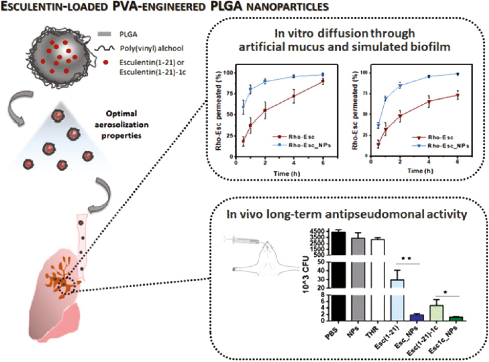
Delivery systems approved by the Food and Drug Administration such as liposomes and poly(lactic-co-glycolic acid) (PLGA) nanoparticles are optimum choices, since they have been approved for use in clinic [39, 40]. Liposomes can encapsulate both hydrophilic and lipophilic molecules including AMPs [41]. He et al. encapsulated polymyxin B (PMB) in liposomes comprising 1,2-dipalmitoyl-sn-glycero-3-phosphocholine and cholesterol [42]. The treatment of liposomal PMB showed a 2-fold lower bacterial burden in lung and longer survival time than free PMB in a pneumonia model. In another study, PEGylated phospholipid (DSPE-PEG2000) was employed to deliver a series of aurein-derived AMPs, peptide 73, D-73, RI-73, and 73c (Figure 1) [43]. The nanocomplex of AMPs and DSPE-PEG2000 showed reduced hemolytic activity and superior antibacterial activity, reducing abscess sizes by more than 60% and lowering bacterial loads up to more than 9-fold over free peptide. Casciaro et al. encapsulated a frog-skin derivative AMP, namely Esc peptides (GIFSKLAGKKIKNLLISGLKG-NH2), in PLGA nanoparticles, which were stabilized with poly(vinyl alcohol) (Figure 2) [44]. As compared with free peptide, Esc peptide-loaded nanoparticles exhibited 2- and 1-fold more penetration through the artificial mucus and simulated biofilm, respectively, and effectively reduced the bacterial loads by more than 4 times in acute lung infection caused by Pseudomonas aeruginosa (P. aeruginosa).
Figure 1 PEGylated phospholipid (DSPE-PEG2000) for the effective delivery of AMPs. The nanocomplex of DSPE-PEG2000 and AMPs showed a low hemolytic activity and high antibacterial efficacy against MRSA in vivo [43]. Reproduced with permission from ACS Infectious Diseases, 2018.
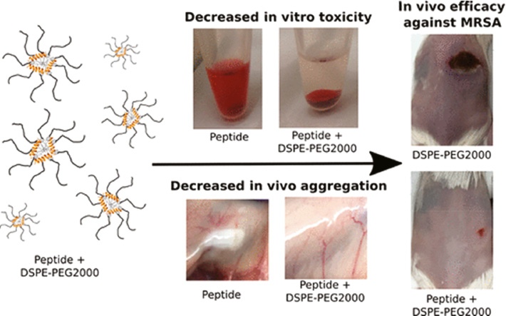
Figure 2 PLGA nanoparticles for the delivery of Esc-derived AMPs. Esc peptide-loaded NPs exhibited more penetration through the artificial mucus and simulated biofilm and effectively reduced the bacterial loads than free peptide [44]. Reproduced with permission from Biomacromolecules, 2019.
Because of the cationic feature of AMPs, anionic nanoparticles have been developed to encapsulate AMPs through electrostatic interaction [24, 45]. Chiara et al. developed a nanosystem (M33-NS) comprising negatively charged dextran nanoparticles and cationic antimicrobial peptide SET-M33 ((KKIRVRLSA)4K2KβA-OH) [46]. M33-NS showed 12-fold more residence time in lung as compared with free peptide, thus improving antimicrobial efficacy against P. aeruginosa in pneumonia model.
Responsive nanoparticles for the delivery of AMPs
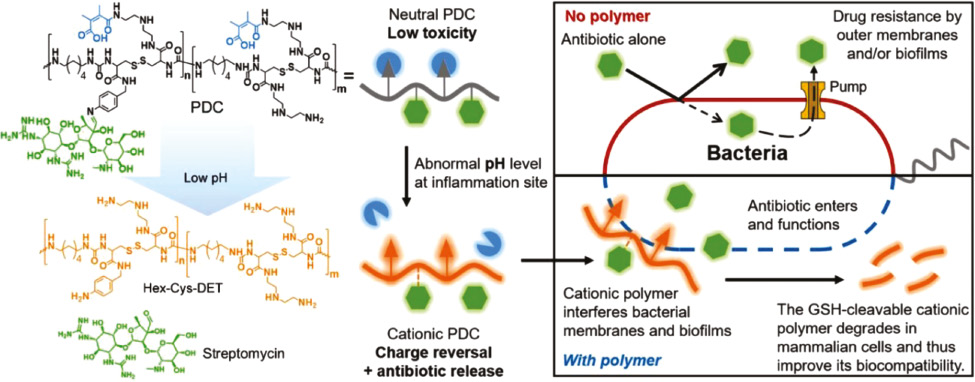
In addition, researchers attempt to selectively deliver and release AMPs by designing infection-responsive nanoparticles [47]. When these nanoparticles reach infected sites, AMPs can be selectively released/activated to inhibit bacterial growth. It has been reported that bacterial infections lead to a decrease of pH as well as an increase of enzymes and reactive oxygen species, forming a special microenvironment [48]. For instance, in consideration of the acidic environment of bacterial infection, Chai et al. designed a pH-sensitive charge reversable polyion nanocomposite (CS-DA) for the delivery of PMB (Figure 3) [47]. CS-DA was comprised of chitooligosaccharides (CS) grafted with 2,3-dimethyl maleic anhydride (DA). The amide bonds, generated from the reaction of DA and amino groups of CS, could be hydrolyzed under acidic microenvironment of bacterial infection. CS-DA could effectively encapsulate PMB through electrostatic interaction in physiological condition. In the acidic infectious environment, the amide bonds were cleaved and converted into positively charged amines, leading to a release of PMB. This design not merely maintained the original excellent bactericidal performance of PMB but also significantly decreased the side effects of PMB such as nephrotoxicity. Acid-cleavable groups can also be used to modify AMPs as well as AMPs analogues except for modifying delivery systems. Ye et al. reported a pH-responsive polymer–drug conjugate (PDC) to fight with antimicrobial-resistant (AMR) pathogens (Figure 4) [49]. The PDC was composed of a polymer Hex-Cys-DET, which was modified with acid-responsive DA groups, and conjugated with streptomycin through a pH-sensitive imine bond. PDC was electrically neutral in the bloodstream due to the modification of DA, showing low toxicity in normal tissues, whereas in the acidic infectious site, PDC transformed to a cationic polymer after cleavage of DA; meanwhile, streptomycin was released, showing a synergistic effect to kill bacteria. The PDC showed higher antimicrobial activity than free streptomycin, causing an average of three orders of reductions of bacteria in three infection models. In a P. aeruginosa-infected lung model, all mice treated with PDC survived, whereas the survival rate of mice was only 40% for free streptomycin group after 4 days. Furthermore, PDC treatment caused negligible toxicity in the major organs.
Figure 3 (A) Composition of CS-DA/PMB nanocomplexes. (B) Charge reversal of CS-DA [47]. Reproduced with permission from Advanced Healthcare Materials, 2020.
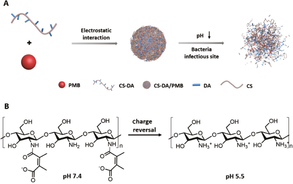
Figure 4 Antibacterial process of the pH-sensitive PDC. The PDC was composed of a polymer Hex-Cys-DET modified with acid-responsive DA groups and conjugated with streptomycin through a pH-sensitive imine bond. In the acidic infectious condition, Hex-Cys-DET transformed to a cationic polymer and streptomycin was released, showing a synergistic effect in reducing bacteria [49]. Reproduced with permission from Advanced Functional Materials, 2020.
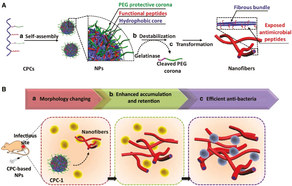
Many enzymes overexpresses in the microenvironment of bacterial infection, such as phospholipase [50], protease lipase [51], and gelatinases [52]. Since they could accelerate the hydrolysis of specific bonds, they inspire researchers to design enzyme-responsive delivery system. Wang et al. developed a polymer–peptide conjugate CPC-1, consisting of a chitosan backbone, an AMP (KLAK), and an enzyme-cleavable peptide (GPL-GVRGC) with a PEG (Figure 5) [53]. CPC-1 assembled into PEGylated nanoparticles, which shielded AMPs and significantly reduced the toxicity of AMPs. CPC-1 showed a significantly decreased hemolytic activity against red blood cells (9.4%) in respect to KLAK treatment (94.3%). Once it reached infectious site, the enzyme-cleavable peptide was cleaved by gelatinase to peel off the protective PEG layer, which led to a transformation of nanoparticles into fibrous nanostructures and exposed AMP (KLAL) to kill bacteria. Interestingly, the in situ morphological transformation significantly increased the accumulation and retention of AMPs in the infected area. CPC-1 showed a long half-life in vivo up to 4 days and high antibacterial activity with a 10-fold decrease of bacterial colony as compared with CPC-2, a control nanoparticle with non-cleavable peptide.
Figure 5 (A) Self-assembly and enzyme-responsive morphology transformation of chitosan–peptide conjugates (CPCs). (a) CPC-1 self-assembled into PEGylated nanoparticles (NPs); (b) bacterial gelatinase cleaved the enzyme-responsive peptide to strip off the protective corona; (c) CPCs transformed into fibrous structures. (B) In situ morphology transformation of CPC to improve the antibacterial efficacy in the infectious sites [53]. Reproduced with permission from Advanced Materials, 2017.
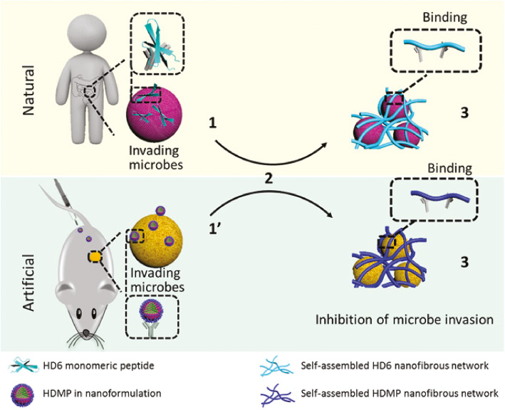
Nanomaterials not only can be designed to target infectious tissue but also can specifically recognize and kill bacteria. Wang et al. designed a human defensin-6 mimic peptide (HDMP), which was constitutive of a ligand peptide (RLYLRIGRR), a β-sheet peptide (KLVFF), and bis-pyrenes (Figure 6) [54]. HDMP self-assembled into sphere nanoparticles with minimal toxicity against normal tissues/cells. At the infection sites, the ligand peptide recognized and specifically bound to lipoteichoic acid on the Gram-positive bacteria to induce a transformation of HDMP nanoparticle into nanofibers. HDMP (5 mg/kg) showed a superior therapeutic effect than vancomycin (5 mg/kg) on the MRSA (methicillin-resistant Staphylococcus aureus)–infected mouse model. All the mice treated by HDMP were alive, whereas the survival rate of the vancomycin group was 83.3% in 96 h.
Figure 6 Antimicrobial process of natural human defensin 6 (HD6) and artificial HD6 mimic peptide (HDMP). The HDMP imitates the HD6 that (2) self-assembles when it encounters microbes, thus (3) forming fibrous networks for capturing invading bacteria [54]. Reproduced with permission from Science Advances, 2020.
Conclusion
Nanoparticle delivery systems have shown great potential for the delivery of AMPs to combat drug-resistant bacteria, particularly in deep organ infections. This strategy provides many advantages over free peptides: it reduces toxicity to normal tissues; it improves the pharmacokinetics of AMPs; it can achieve superior distribution of AMPs in the infection tissues with enhanced bioavailability; it can be designed as intelligent nanocarriers for target infectious tissues and bacteria. In the future, the development goal of the delivery systems of AMPs is to design AMPs delivery systems to achieve selective activation and active target in the infectious sites, thus improving the therapeutic efficacy against bacterial infection and reducing the toxicity against normal tissues.
Conflicts of interest
There are no conflicts to declare.
Acknowledgements
This work was supported by the National Natural Science Foundation of China (51873070, U1801252 and 82071860), the Key Research and Development Program of Guangzhou (202007020002), and the Natural Science Foundation of Guangdong Province (2018A030313110).
References
- World Health Organization News Release. Antimicrobial resistance 2020. Available from: https://www.who.int/news-room/fact-sheets/detail/antimicrobial-resistance. [Last accessed on 30 Dec 2020].
- Song M, Liu Y, Huang X, Ding S, Wang Y, et al. A broad-spectrum antibiotic adjuvant reverses multidrug-resistant Gram-negative pathogens. Nat Microbiol 2020;5:1040-50. [PMID: 32424338 DOI: 10.1038/s41564-020-0723-z]
- Li S, Dong S, Xu W, Tu S, Yan L, et al. Antibacterial hydrogels. Adv Sci (Weinh) 2018;5:1700527. [PMID: 29876202 DOI: 10.1002/advs.201700527]
- Mookherjee N, Anderson MA, Haagsman HP, Davidson D. Antimicrobial host defence peptides: functions and clinical potential. Nat Rev Drug Discov 2020;19:311-32. [PMID: 32107480 DOI: 10.1038/s41573-019-0058-8]
- Wang G, Li X, Wang Z. APD3: the antimicrobial peptide database as a tool for research and education. Nucleic Acids Res 2016;44:D1087-93. [PMID: 26602694 DOI: 10.1093/nar/gkv1278]
- Lazzaro BP, Zasloff M, Rolff J. Antimicrobial peptides: application informed by evolution. Science 2020;368:eaau5480. [PMID: 32355003 DOI: 10.1126/science.aau5480]
- Brogden KA. Antimicrobial peptides: pore formers or metabolic inhibitors in bacteria? Nat Rev Microbiol 2005;3:238-50. [PMID: 15703760 DOI: 10.1038/nrmicro1098]
- Thapa RK, Diep DB, Tønnesen HH. Topical antimicrobial peptide formulations for wound healing: current developments and future prospects. Acta Biomater 2020;103:52-67. [PMID: 31874224 DOI: 10.1016/j.actbio.2019.12.025]
- Fjell CD, Hiss JA, Hancock RE, Schneider G. Designing antimicrobial peptides: form follows function. Nat Rev Drug Discov 2011;11:37-51. [PMID: 22173434 DOI: 10.1038/nrd3591]
- Ciumac D, Gong H, Hu X, Lu JR. Membrane targeting cationic antimicrobial peptides. J Colloid Interface Sci 2019;537:163-85. [PMID: 30439615 DOI: 10.1016/j.jcis.2018.10.103]
- Guan Q, Huang S, Jin Y, Campagne R, Alezra V, et al. Recent advances in the exploration of therapeutic analogues of gramicidin S, an old but still potent antimicrobial peptide. J Med Chem 2019;62:7603-17. [PMID: 30938996 DOI: 10.1021/acs.jmedchem.9b00156]
- Carratalá JV, Serna N, Villaverde A, Vázquez E, Ferrer-Miralles N. Nanostructured antimicrobial peptides: the last push towards clinics. Biotechnol Adv 2020;44:107603. [PMID: 32738381 DOI: 10.1016/j.biotechadv.2020.107603]
- Costa ML, Achten J, Knight R, Bruce J, Dutton SJ, et al. Effect of incisional negative pressure wound therapy vs standard wound dressing on deep surgical site infection after surgery for lower limb fractures associated with major trauma: the WHIST randomized clinical trial. JAMA 2020;323:519-26. [PMID: 32044942 DOI: 10.1001/jama.2020.0059]
- Mahlapuu M, Håkansson J, Ringstad L, Björn C. Antimicrobial peptides: an emerging category of therapeutic agents. Front Cell Infect Microbiol 2016;6:194-206. [PMID: 28083516 DOI: 10.3389/fcimb.2016.00194]
- Schluesener H, Radermacher S, Melms A, Jung S. Leukocytic antimicrobial peptides kill autoimmune T cells. J Neuroimmunol 1993;47:199-202. [PMID: 8370771 DOI: 10.1016/0165-5728(93)90030-3]
- Trimble MJ, Mlynárčik P, Kolář M, Hancock RE. Polymyxin: alternative mechanisms of action and resistance. Cold Spring Harb Perspect Med 2016;6:a025288. [PMID: 27503996 DOI: 10.1101/cshperspect.a025288]
- Hancock RE, Sahl HG. Antimicrobial and host-defense peptides as new anti-infective therapeutic strategies. Nat Biotechnol 2006;24:1551-7. [PMID: 17160061 DOI: 10.1038/nbt1267]
- Borro BC, Malmsten M. Complexation between antimicrobial peptides and polyelectrolytes. Adv Colloid Interface Sci 2019;270:251-60. [PMID: 31301601 DOI: 10.1016/j.cis.2019.07.001]
- Nordström R, Malmsten M. Delivery systems for antimicrobial peptides. Adv Colloid Interface Sci 2017;242:17-34. [PMID: 28159168 DOI: 10.1016/j.cis.2017.01.005]
- Pelaz B, Alexiou C, Alvarez-Puebla RA, Alves F, Andrews AM, et al. Diverse applications of nanomedicine. ACS Nano 2017;11:2313-81. [PMID: 28290206 DOI: 10.1021/acsnano.6b06040]
- Sadikot RT. The potential role of nano- and micro-technology in the management of critical illnesses. Adv Drug Deliv Rev 2014;77:27-31. [PMID: 25204519 DOI: 10.1016/j.addr.2014.07.004]
- Mitragotri S, Anderson DG, Chen X, Chow EK, Ho D, et al. Accelerating the translation of nanomaterials in biomedicine. ACS Nano 2015;9:6644-54. [PMID: 26115196 DOI: 10.1021/acsnano.5b03569
- Water JJ, Kim Y, Maltesen MJ, Franzyk H, Foged C, et al. Hyaluronic acid-based nanogels produced by microfluidics-facilitated self-assembly improves the safety profile of the cationic host defense peptide novicidin. Pharm Res 2015;32:2727-35. [PMID: 25813840 DOI: 10.1007/s11095-015-1658-6]
- Nordström R, Andrén OCJ, Singh S, Malkoch M, Davoudi M, et al. Degradable dendritic nanogels as carriers for antimicrobial peptides. J Colloid Interface Sci 2019;554:592-602. [PMID: 31330426 DOI: 10.1016/j.jcis.2019.07.028]
- Fumakia M, Ho EA. Nanoparticles encapsulated with LL37 and serpin A1 promotes wound healing and synergistically enhances antibacterial activity. Mol Pharm 2016;13:2318-31. [PMID: 27182713 DOI: 10.1021/acs.molpharmaceut.6b00099]
- Yuan D, Zhao Y, Banks WA, Bullock KM, Haney M, et al. Macrophage exosomes as natural nanocarriers for protein delivery to inflamed brain. Biomaterials 2017;142:1-12. [PMID: 28715655 DOI: 10.1016/j.biomaterials.2017.07.011]
- Qin X, Yu C, Wei J, Li L, Zhang C, et al. Rational design of nanocarriers for intracellular protein delivery. Adv Mater 2019;31:e1902791. [PMID: 31496027 DOI: 10.1002/adma.201902791]
- Li B, Zhang X, Dong Y. Nanoscale platforms for messenger RNA delivery. Wiley Interdiscip Rev Nanomed Nanobiotechnol 2019;11:e1530. [PMID: 29726120 DOI: 10.1002/wnan.1530]
- Jasinski D, Haque F, Binzel DW, Guo P. Advancement of the emerging field of RNA nanotechnology. ACS Nano 2017;11:1142-64. [PMID: 28045501 DOI: 10.1021/acsnano.6b05737]
- Scaletti F, Hardie J, Lee YW, Luther DC, Ray M, et al. Protein delivery into cells using inorganic nanoparticle-protein supramolecular assemblies. Chem Soc Rev 2018;47:3421-32. [PMID: 29537040 DOI: 10.1039/c8cs00008e]
- Kopp M, Kollenda S, Epple M. Nanoparticle–protein interactions: therapeutic approaches and supramolecular chemistry. Acc Chem Res 2017;50:1383-90. [PMID: 28480714 DOI: 10.1021/acs.accounts.7b00051]
- Giodini L, Re FL, Campagnol D, Marangon E, Posocco B, et al. Nanocarriers in cancer clinical practice: a pharmacokinetic issue. Nanomedicine 2017;13:583-99. [PMID: 27520727 DOI: 10.1016/j.nano.2016.07.012]
- Reinholz J, Landfester K, Mailänder V. The challenges of oral drug delivery via nanocarriers. Drug Deliv 2018;25:1694-705. [PMID: 30394120 DOI: 10.1080/10717544.2018.1501119]
- Suchaoin W, Bernkop-Schnürch A. Nanocarriers protecting toward an intestinal pre-uptake metabolism. Nanomedicine 2017;12:255-69. [PMID: 28093952 DOI: 10.2217/nnm-2016-0331]
- Farokhzad OC, Langer R. Impact of nanotechnology on drug delivery. ACS Nano 2009;3:16-20. [PMID: 19206243 DOI: 10.1021/nn900002m]
- Foulkes R, Man E, Thind J, Yeung S, Joy A, et al. The regulation of nanomaterials and nanomedicines for clinical application: current and future perspectives. Biomater Sci 2020;8:4653-64. [PMID: 32672255 DOI: 10.1039/d0bm00558d]
- Saw PE, Jon S. Understanding of the entry mechanism of nanoparticles into tumors determines the future direction of nanomedicine development. BIO Integration 2020;1:193-5. [DOI: 10.15212/bioi-2020-0033]
- Azzopardi EA, Ferguson EL, Thomas DW. The enhanced permeability retention effect: a new paradigm for drug targeting in infection. J Antimicrob Chemother 2013;68:257-74. [PMID: 23054997 DOI: 10.1093/jac/dks379]
- Fan Y, Zhang Q. Development of liposomal formulations: from concept to clinical investigations. Asian J Pharm Sci 2013;8:81-7. [DOI: 10.1016/j.ajps.2013.07.010]
- Li X, Jiang X. Microfluidics for producing poly (lactic-co-glycolic acid)-based pharmaceutical nanoparticles. Adv Drug Deliv Rev 2018;128:101-14. [PMID: 29277543 DOI: 10.1016/j.addr.2017.12.015]
- Lee Y, Thompson DH. Stimuli-responsive liposomes for drug delivery. Wiley Interdiscip Rev Nanomed Nanobiotechnol 2017;9:e1450. [PMID: 28198148 DOI: 10.1002/wnan.1450]
- He J, Abdelraouf K, Ledesma KR, Chow DS-L, Tam VH. Pharmacokinetics and efficacy of liposomal polymyxin B in a murine pneumonia model. Int J Antimicrob Agents 2013;42:559-64. [PMID: 24016799 DOI: 10.1016/j.ijantimicag.2013.07.009]
- Kumar P, Pletzer D, Haney EF, Rahanjam N, Cheng JT, et al. Aurein-derived antimicrobial peptides formulated with pegylated phospholipid micelles to target methicillin-resistant Staphylococcus aureus skin infections. ACS Infect Dis 2018;5:443-53. [PMID: 30565465 DOI: 10.1021/acsinfecdis.8b00319]
- Casciaro B, d’Angelo I, Zhang X, Loffredo MR, Conte G, et al. Poly (lactide-co-glycolide) nanoparticles for prolonged therapeutic efficacy of esculentin-1a-derived antimicrobial peptides against Pseudomonas aeruginosa lung infection: in vitro and in vivo studies. Biomacromolecules 2019;20:1876-88. [PMID: 31013061 DOI: 10.1021/acs.biomac.8b01829]
- Gao J, Wang M, Wang F, Du J. Synthesis and mechanism insight of a peptide-grafted hyperbranched polymer nanosheet with weak positive charges but excellent intrinsically antibacterial efficacy. Biomacromolecules 2016;17:2080-6. [PMID: 27181113 DOI: 10.1021/acs.biomac.6b00307]
- Falciani C, Zevolini F, Brunetti J, Riolo G, Gracia R, et al. Antimicrobial peptide-loaded nanoparticles as inhalation therapy for Pseudomonas aeruginosa infections. Int J Nanomed 2020;15:1117-28. [PMID: 32110011 DOI: 10.2147/IJN.S218966]
- Chai M, Gao Y, Liu J, Deng Y, Hu D, et al. Polymyxin B-polysaccharide polyion nanocomplex with improved biocompatibility and unaffected antibacterial activity for acute lung infection management. Adv Healthc Mater 2020;9:1901542. [PMID: 31898875 DOI: 10.1002/adhm.201901542]
- Yamamoto S, Yamazaki S, Shimizu T, Takeshima T, Fukuma S, et al. Body temperature at the emergency department as a predictor of mortality in patients with bacterial infection. Medicine 2016;95:e3628. [PMID: 27227924 DOI: 10.1097/MD.0000000000003628]
- Ye M, Zhao Y, Wang Y, Yodsanit N, Xie R, et al. pH-Responsive polymer–drug conjugate: an effective strategy to combat the antimicrobial resistance. Adv Funct Mater 2020;30:2002655. [DOI: 10.1002/adfm.202002655]
- Xiong MH, Li YJ, Bao Y, Yang XZ, Hu B, et al. Bacteria-responsive multifunctional nanogel for targeted antibiotic delivery. Adv Mater 2012;24:6175-80. [PMID:22961974 DOI: 10.1002/adma.201202847
- Rosenau F, Jaeger K-E. Bacterial lipases from Pseudomonas: regulation of gene expression and mechanisms of secretion. Biochimie 2000;82:1023-32. [PMID: 11099799 DOI: 10.1016/s0300-9084(00)01182-2]
- Lin A, Liu Y, Zhu X, Chen X, Liu J, et al. Bacteria-responsive biomimetic selenium nanosystem for multidrug-resistant bacterial infection detection and inhibition. ACS Nano 2019;13:13965-84. [PMID: 31730327 DOI: 10.1021/acsnano.9b05766]
- Qi G-B, Zhang D, Liu F-H, Qiao Z-Y, Wang H. An “on-site transformation” strategy for treatment of bacterial infection. Adv Mater 2017;29:1703461. [PMID: 28782856 DOI: 10.1002/adma.201703461]
- Fan Y, Li XD, He PP, Hu XX, Zhang K, et al. A biomimetic peptide recognizes and traps bacteria in vivo as human defensin-6. Sci Adv 2020;6:eaaz4767. [PMID: 32494712 DOI: 10.1126/sciadv.aaz4767]