Copper-Based Nanomaterials for Image-Guided Cancer Therapy
1Key Laboratory of Medical Imaging Precision Theranostics and Radiation Protection, University of South China, College of Hunan Province, Changsha, Hunan 410004, China
2Institute of Medical Imaging, Hengyang Medical School, University of South China, Hengyang, Hunan, 421001 China
3The Seventh Affiliated Hospital, Hunan Veterans Administration Hospital, Hengyang Medical School, University of South China, Changsha, Hunan, China
4School of Dentistry, Federal University of Ceará, Sobral, Ceará, Brazil
5School of Medicine, Federal University of Ceará, Sobral, Ceará, Brazil
*Correspondence to: Meng Du, E-mail: dumeng_work@126.com; Mirna Marques Bezerra, E-mail: mirna@ufc.br
Received: 3 April 2024; Revised: 7 June 2024; Accepted: 11 June 2024; Published Online: August 1 2024
Cite this paper:
Xu H, Guo Z, Li M et al. Copper-Based Nanomaterials for Image-Guided Cancer Therapy. BIO Integration 2024; 5: 1–14.
DOI: 10.15212/bioi-2024-0013. Available at: https://bio-integration.org/
Download citation
© 2024 The Authors. This is an open access article distributed under the terms of the Creative Commons Attribution License (https://creativecommons.org/licenses/by/4.0/). See https://bio-integration.org/copyright-and-permissions/
Abstract
Cancer is a significant disease that poses a major threat to human health. Image-guided cancer therapy refers to a series of medical procedures that use imaging technology to precisely locate and treat cancer. Combining the dual characteristics of medical images and functional nanomaterial (NM) drug carriers, various integrated diagnosis and treatment probes have been developed for in vivo dynamic monitoring and therapeutic effect evaluation of drugs based on medical imaging. Copper (Cu)-based NMs have emerged as valuable products of nanotechnology due to their unique physicochemical properties, which are influenced by factors, such as size, shape, and surface properties. In the field of imaging, Cu-based NMs offer a combination of desirable characteristics, including fluorescence emission, contrast enhancement, and radiolabeling stability. These properties form the foundation for a wide range of imaging modalities. In addition, Cu-based NMs can be used as a carrier for diagnostic or therapeutic drugs and the synergistic effect of multiple therapeutic modalities can be realized by doping multiple transition metals into the heterostructures. These properties have become an important basis for imaging-guided therapy with Cu-based NMs. In this review we introduce biocompatible Cu-based NMs for image-guided cancer therapy and provide an overview of the promising outcomes in biomedical research.
Keywords
Copper-based nanomaterials, image-guided, imaging properties, multimodal imaging.
Introduction
Cancer poses a significant threat to human life and health. The primary clinical methods used for treating cancer include surgery, radiotherapy, and chemotherapy. However, these treatments often have significant drawbacks, such as substantial surgical trauma, high toxicity and side effects, incomplete treatment, and strong resistance to drugs. Therefore, developing cancer treatments that are specifically lethal to cancer cells, safe, and efficient remains a scientific challenge and a direction for future research [1]. Image-guided therapy, which combines imaging and therapeutic functions, holds great potential for enhancing the effectiveness of anticancer treatments, while reducing side effects [2]. Traditional cancer treatments generally involve surgery, which directly affects tumors and is the preferred treatment for most cancers. However, surgery poses high risks and causes significant trauma to the human body. Surgery conducted with the aid of imaging guidance through minimally invasive techniques, such as punctures and catheters, greatly alleviates patient suffering while treating the tumor. Radiotherapy and chemotherapy can effectively stop the proliferation, infiltration, and metastasis of cancer cells, but lack specificity in distinguishing between cancerous and normal cells. Consequently, radiotherapy and chemotherapy inevitably damage healthy cells while killing cancer cells, leading to severe side effects on healthy tissues and organs. Utilizing image guidance for radiotherapy and chemotherapy can precisely locate tumors, ensuring treatment accuracy. Real-time monitoring of treatment effects based on imaging feedback allows for timely adjustments to the treatment plan. This approach offers personalized treatment options for cancer patients, enhancing the overall efficiency of cancer treatments [3].
Imaging-guided therapy represents a synergistic approach that combines diagnosis and therapy in the field of nanomedicine, which is known as theranostics. This approach has the potential to offer more personalized therapeutic strategies. The exploration of innovative nanoplatforms that integrate diagnostics and therapeutics has garnered significant interest in improving tumor diagnosis and treatment. For example, methodologies of photonic clustered regularly interspaced short palindromic repeat (CRISPR) sensing (MOPCS), which combines an optical sensing technology-surface plasmon resonance (SPR) with a ‘gene scissors’ CRISPR technique, has achieved both high sensitivity and specificity measurement [4–6]. Traditional diagnostic-integrated materials involve the integration of diagnostic contrast agents and antitumor drugs within the same nanocarrier, thereby creating a multifunctional diagnostic platform that encompasses diagnostic, therapeutic, and efficacy monitoring functions [7]. However, in practical applications, the complex preparation process and the difficulty of synergizing imaging and therapeutic functions make it difficult to translate the diagnostic-therapeutic integrated nanoplatforms to the clinic [8]. Transition metal element nanosystems are one of the most representative nanomaterials in nanomedicine, and copper (Cu)-based nanomaterials (NMs) have high therapeutic and diagnostic performance in biomedicine due to easily tunable nanostructures and compositions, as well as unique physicochemical properties and biological effects [9].
Cu-based NMs have garnered significant attention due to advantages over silver or gold counterparts. The benefits of Cu-based NMs include the high yield achieved through mild synthetic conditions and Cu abundance and low cost, which facilitate practical applications in large-scale nanotechnology and affordable healthcare settings. Notably, Cu is an essential trace element in the human body that allows for effective removal through physiologic processes when present in excess [10]. This characteristic provides Cu-based NMs with an additional advantage in diagnostic and therapeutic applications. Cu-based NMs offer greater control and tunability over their structure, composition, and size during the preparation process compared to other NMs, thereby expanding their potential for diverse biomedical applications [11]. This review presents an overview of the development of image-guided cancer therapy, summarizes the imaging properties of Cu-based NMs, and discusses the application of Cu-based NMs in image-guided visualization of cancer therapy in diagnostics and therapeutics.
Current status of image-guided cancer therapy
Advanced imaging techniques provide valuable information on disease stage, location, and aggressiveness, which enable the implementation of effective and definitive treatment strategies. Real-time image guidance has a pivotal role in delivering “precision therapy” across various treatment modalities, such as surgery, radiotherapy, and chemotherapy. Lesions can be precisely targeted and eliminated or eradicated by utilizing image-guided real-time “precision therapy,” which minimizes harm to adjacent healthy tissue [12]. Image-guided surgery (IGS) holds promise for achieving individualized precision surgery by optimizing resection accuracy and striking a balance between radicality and side effects [13]. Imaging has always been integral to treatment planning and delivery in radiation oncology. Recent advances in imaging have significantly improved treatment efficacy and reduced toxicity with ongoing developments expected [14]. Utilizing imaging to track pharmacokinetics, monitor chemotherapy processes and effects, and visualize or quantify drug delivery systems has become feasible, thus offering opportunities to enhance therapeutic outcomes in a personalized manner [15].
The rapid development of nanoscience has introduced new strategies for cancer treatment compared to general image-guided therapy. Nanomedicine enables personalized/precision medicine to precisely locate lesions, monitor responses, maximize therapeutic efficiency, and verify treatment effectiveness, thereby significantly reducing costs throughout the medical process. In recent years there has been a growing interest in utilizing theranostic nanomedicine for image-guided therapy. Nanoparticles (NPs) designed for therapy possess inherent imaging capabilities, which are harnessed to guide cancer treatment. These nanoformulations are often engineered to incorporate contrast agents, enabling visualization of the cancer therapy process [16]. Theranostics has gained considerable recognition for its capacity to integrate real-time cancer diagnosis with effective treatment strategies. By covalently conjugating imaging agents, therapeutic agents, stimuli-responsive linkers, and/or targeting molecules, it is possible to synthesize activatable multifunctional molecular agents. These agents can be selectively triggered in tumor sites by overexpressed physiologic stimuli or external triggers, resulting in the release of imaging agents and cytotoxic drugs. This approach presents multiple benefits for tumor imaging and therapy, encompassing a high signal-to-noise ratio, minimal systemic toxicity, and enhanced therapeutic effects [17].
However, there are still some limitations to theranostics include technical complexity and potential risks. Specifically, the time and spatial resolution of imaging equipment is difficult to meet the diagnostic accuracy requirements and the in vivo circulation time and tumor targeting of biological NMs cannot meet the long-term safety requirements. Cu nanostructures have the following important advantages compared to other NMs: i) synthesis of nanostructures usually has adjustable size, morphology, and surface physical and chemical properties; ii) nanocarriers can be used as diagnostic or therapeutic agents and used to build more functional diagnostic probes; and iii) numerous transition metals can be doped into heterostructures, showing functional synergies [18]. Therefore, Cu-based NMs can solve the above problems and are one of the most widely used NMs at present.
Imaging properties of copper-based nanomaterials
The imaging properties of Cu-based NMs include fluorescence (FL) emission, contrast enhancement, and radiolabeling stability, which are closely related to image imaging performance (Figure 1).
Figure 1 Copper-based nanomaterials characterization and imaging advantages. (FL, fluorescence; PAI, photoacoustic imaging; MRI, magnetic resonance imaging; US, ultrasound; PET, positron emission tomography).
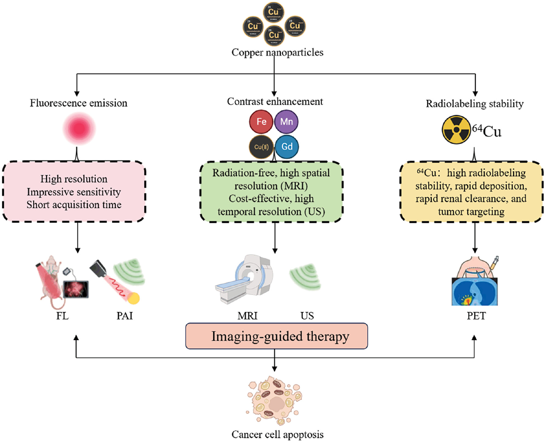
Fluorescence emission
FL emission refers to the energy release that occurs when electrons transition between two energy states within a luminescent material. When metal NPs attain sizes comparable to the Fermi wavelength of electrons, the NPs exhibit discrete energy levels. This characteristic gives rise to intriguing physical and chemical properties, including strong FL. Cu is a readily accessible and cost-effective metal compared to noble metals. Theoretical and experimental studies have confirmed that Cu-based NMs have the capability to absorb photon energy when exposed to laser radiation, leading to electron transitions. During the relaxation of excited electrons to the ground state, various energy transfer pathways arise, resulting in Cu-based NMs possessing distinctive photoluminescent properties [19].
FL imaging, particularly in the near-infrared (NIR) range, offers high-resolution images with remarkable sensitivity and fast acquisition times, making NIR a promising tool for real-time biomolecule detection and tumor diagnosis [20]. An important study by Meng et al. [21] conducted a significant study that focused on the development of a DNA@Cu-metal organic framework (MOF) nanosystem tailored for FL imaging of human breast cancer. This nanosystem spontaneously degrades after entering hypoxic tumors and achieves efficient Cu(II)-dependent DNAzyme signal amplification. The intensity of the Cu(II)-induced carboxyfluorescein (FAM) green FL signal was shown to be nearly 3.49-fold higher than other metal ions, which shows that this nanosystem has a promising application in FL imaging of hypoxic cancers. In the area of cellular imaging, Chowdhury et al. [22] rationally designed double-stranded Deoxyribonucleic acid (DNA) oligonucleotides with two cholesterols that spontaneously form the lipid-mediated DNA micelles and generate a high fluorescence signal after the formation of DNA-templated Copper nanocluster (CuNCs). The cell membranes of Mucin 1 (MUC1)-positive cancer cells with well-defined DNA nanostructures are stained by CuNCs that exhibit an intense, red FL signal that is clearly distinguished from MUC1-negative cancer cells.
Contrast enhancement
Cu-based NMs have been effectively synthesized as contrast agents for use in an array of cancer imaging modalities [23]. Magnetic resonance imaging (MRI) is a technique that involves the alignment and relaxation of hydrogen protons within an external magnetic field. MRI excels as a structural imaging modality that provides high-resolution images, especially of soft tissues. The sensitivity of MRI has been greatly enhanced by the introduction of nanoscale contrast agents that can amplify the detection sensitivity by several orders of magnitude. This advance has enabled the traditionally macroscopic imaging method to discern unique molecular signatures [24]. In addition to the commonly used ferrum (Fe)-, manganese (Mn)-, and gadolinium (Gd)-based magnetic NPs (MNPs), Cu(II) has demonstrated efficacy as a potent contrast agent in MRI [25]. For example, CuS NPs with a high-activity surface have shown the potential for pH and NIR light-responsive T1-weighted MRI, especially in detecting breast cancer [BC] [26].
Multimodal medical imaging is gaining traction clinically because multimodal medical imaging allows for the integration of data from different physical phenomena, which provides a more comprehensive understanding of pathologic conditions. Nano-sized contrast agents have a crucial role in this advance, potentially boosting the sensitivity of each imaging modality and enabling precise tumor visualization [27]. CuO NPs are emerging as promising candidates for combined MR-ultrasound (US) imaging. CuO NPs offer radiation-free MRI scans with high spatial resolution as well as cost-effective US examinations with high temporal resolution. Studies have shown that CuO NPs effectively shorten the magnetic T1 relaxation time, while also influencing the speed of sound and the ultrasonic attenuation coefficient. Notably, these effects are concentration-dependent and when the NP concentration reaches a specific level, a clearly visible and quantifiable contrast enhancement effect is observed [27].
Radiolabeling stability
Cu radionuclides, specifically 64Cu, possess favorable nuclear decay properties that make Cu radionuclides suitable for utilization in nuclear medicine applications [28]. NMs labeled with 64Cu exhibit noteworthy attributes, including high stability of radiolabeling, accelerated deposition, rapid renal clearance, and effective targeting of tumors. Positron emission tomography (PET), a widely used nuclear imaging technique in clinical practice, is known for exceptional sensitivity and almost unlimited tissue penetration depth [29]. In the radioactive form, Cu emits positrons suitable for PET imaging of cancerous tissues. Remarkably, radioactive Cu can be incorporated directly into Cu sulfide NPs, eliminating the need for a radiometal chelator. Cui et al. [30] crafted a series of Cu sulfide NPs integrated with radioactive Cu. These NPs served as a PET contrast agent, facilitating a study of the application in tumor detection and biological metabolism analysis. When peg-coated radioactive Cu-labeled Cu sulfide NPs were administered to tumor-bearing mice, the tumors exhibited significant uptake, providing an excellent imaging effect.
Gao et al. [31] utilized pre-conjugation of luteinizing hormone-releasing hormone (LHRH) to bovine serum albumin (BSA) scaffolds to construct [64Cu] CuNCs@BSA-LHRH for PET imaging in an orthotopic lung cancer model. The nanoclusters were shown to exhibit high radiolabeling stability and were heavily absorbed in A549 tumors and kidneys. Notably, the [64Cu] CuNCs exhibited remarkable features, such as high radiolabel stability, rapid deposition, fast renal elimination, and efficient tumor targeting. PET imaging using [64Cu] CuNCs as a radiolabel is superior to NIR FL imaging, and provides more sensitive, precise, and in-depth in vivo lung cancer imaging [32].
Applications of Cu-based NMs in image-guided cancer therapy
In recent years significant progress has been made in the development of novel cancer therapeutics and treatment strategies, with the goal of extending overall survival and enhancing the quality of life for cancer patients [33,34]. Regulating Cu levels in tumor cells has emerged as a promising approach for cancer treatment [35]. Cu is involved in regulating multiple aspects of the onset and progression of malignant tumors, including growth, angiogenesis, and metastasis [36]. Insufficient Cu affects the biological functions of Cu-binding enzymes, while excess Cu can overload and even kill cells, prompting the development of Cu-specific chelators and Cu ion carriers to control cancer development by lowering or increasing intracellular Cu levels in tumor cells. Blood vessels are sensitive to Cu. In the absence of serum and growth factors, Cu promotes the proliferation of human endothelial cells, whereas the same concentration of zinc or iron slows the growth. Deprivation of Cu can turn off the angiogenesis ‘switch,’ halting endothelial cell proliferation and arresting endothelial cells in the G0 phase. Inhibiting Cu transporters or Cu chaperone proteins, such as CTR1 and ATOX1, can also lead to Cu homeostasis imbalance and achieve anti-angiogenic effects. These findings indicate that Cu is a key element in promoting tumor angiogenesis [37]. LOX and lysyl oxidase-like (LOXL) proteins are involved in the cross-linking of collagen and elastin, with Cu being essential for the activity of both proteins. Blocking the activity of LOX and LOXL through Cu regulation is an effective treatment to inhibit cancer metastasis [38]. Cu-based NMs have unique properties that make Cu-based NMs ideal for antitumor applications, including convenience, efficiency, and safety. BC is the second most common cancer affecting women worldwide. Studies have shown that Cu-based NMs have broad application prospects in the treatment of BC. For example, Ahamed et al. [39] reported that copper ferrite (CuFe2O4) NPs added to the culture of human BC MCF-7 cells can cause an intracellular oxidation stress response, exerting anti-cancer effects. Furthermore, Rajagopal et al. [40] found that copper nanoparticles (Wt-CuNPs) have obvious cytotoxic effects on MCF-7 cells. The specific mechanism is mainly due to the release of Cu ions from the NPs and the binding of Cu ions to tumor cell DNA, thus causing DNA damage and apoptosis. Cu-based NMs have also achieved good results in the treatment of esophageal and lung cancers. Wang et al. [41] coated Cu9S5 NPs with silica to form Cu9S5@MS core-shell nanostructures and added the Cu9S5@MS core-shell nanostructures to human esophageal squamous carcinoma Eca109 and TE8 cells. Cu9S5@MS + NIR showed anticancer activity by arresting cell cycle in EC109 and TE8 cancer cell co-cultures. Naatz et al. [42] showed that doped Fe can be used to control dissolution kinetics of Cu-based NMs using Fe-doped CuO NMs. These particles also trigger a systemic anti-cancer immune response, promote the generation of reactive oxygen species (ROS), and increase the rate of lung squamous cancer cell death. In a detailed preclinical mouse model study, the in vitro normal cell viability and the ex vivo hemolysis assay revealed the biocompatible nature of Cu-based NMs [43]. Cu-based NMs show selective cytotoxicity towards cancer cells in a concentration-dependent manner, while demonstrating minimal toxicity towards normal cells, highlighting the safety profile [44]. Therefore, the utilization of Cu-based NMs for imaging-guided therapy has garnered increasing attention, as depicted in Figure 2. Table 1 provides an overview of the diverse roles of various Cu-based NMs in image-guided tumor therapy.
Figure 2 Copper-based nanomaterials for image-guided cancer therapy. (GSH, reduced glutathione; GSSH, oxidized glutathione).
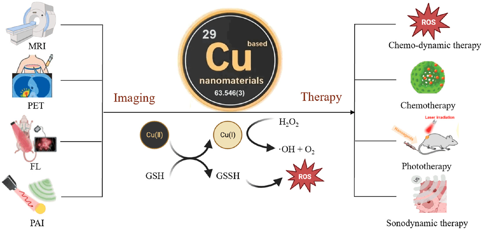
Table 1 Cu-based NMs in Tumor Imaging and Therapy
| Classification | Imaging Modality | Cu-based NMs | Therapy | Ref. |
|---|---|---|---|---|
| Single mode imaging | MR imaging | [64Cu]-CuNCs | FUS | [51] |
| UIONPs | PDT | [54] | ||
| PDA@CFNs | PTT/chemotherapy | [59] | ||
| PET imaging | Cit-[64Cu]CuS NPs | PTT | [68] | |
| PEG-[64Cu]CuS NPs | PTT | [68] | ||
| CuS@MSN NPs | PTT/chemotherapy | [71] | ||
| CuTz-1@F127 MOF | PDT | [72] | ||
| FL imaging | Cu-CDs | PDT | [75] | |
| Cu(II)-CDs NPs | CDT | [83] | ||
| NP-Cu | PDT/PTT/CDT | [92] | ||
| PA imaging | ZnS/Cu2O@ZIF-8@PVP | PTT/CDT | [100] | |
| Gd:CuS@BSA NPs | PTT | [102] | ||
| Cu(II)/LRu/PDA NPs | PDT/PTT | [103] | ||
| Multimodal imaging | PA/MR imaging | Gd:CuS@BSA NPs | PTT | [102] |
| MR/PAT imaging | Cu(II)/LRu/PDA NPs | PDT/PTT | [103] | |
| PA/MR imaging | Cu@PAA NCs | SDT | [109] | |
| UCL/MR/PAT imaging | mUCNPs@DOX/CuS/HA | PTT/Chemotherapy | [111] |
MRI-guided cancer therapy
Tracking the site where the drug reaches and monitoring the real-time effects of tumor therapy are essential for accurate treatment and evaluation, especially in deep tissues [45–47]. To achieve this goal, various imaging modalities are required, among which MRI is a promising and mature technique for deep tissue imaging without the need for radionuclides or ionizing radiation [48,49]. Due to the ability of intracellular H2O2 to oxidize antimagnetic Cu(I) to paramagnetic Cu(II), Cu-based NMs have been developed as contrast agents for MRI and are widely utilized in MRI imaging [50]. Zhang et al. [51] conducted a study utilizing MRI to facilitate the opening of the blood-brain barrier (BBB) in diffuse pontine gliomas through focused ultrasound (FUS). In this study radiolabeled nanoclusters ([64Cu]-CuNCs) were administered and it was observed that the [64Cu]-CuNCs efficiently reached the pontine bridges of naive mice. The volume of radioactive signal distribution within the brain bridges increased from 21.90 ± 2.24 mm3 to 34.4 ± 2.22 mm3 within 24–48 h, indicating the dynamic diffusion and prolonged intra-tumoral retention of [64Cu]-CuNCs in the tumor.
Cu-based NMs can be utilized for photothermal therapy (PTT) and photodynamic therapy (PDT) in conjunction with MRI guidance [52]. PTT and PDT involve the conversion of light energy into heat and the generation of ROS, resulting in cancer cell death. Cu is an essential trace element in the human body, and due to its localized surface plasmon resonance (LSPR) properties, Cu-based NMs have good NIR absorption and excellent photothermal properties. At the same time, Cu (II) is easily reduced to Cu (I) by the Haber-Weiss reaction [Cu (II) + •O2 → Cu (I) + O2] by glutathione (GSH) in cancer cells. This process triggers a Fenton-like reaction [Cu (I) + H2O2 → Cu (II) + OH− + •OH] that relieves tumor hypoxia while generating ROS, which enhances the efficacy of PTT/PDT to synergistically destroy tumor cells [53]. Researchers have developed polyacrylic acid (PAA)-coated ultra-small iron oxide NPs (UIONPs) that effectively encapsulate Cu(II) and were loaded with a NIR dye (IR-780) to enhance PDT for cancer treatment [54]. Additionally, due to the small size (<10 nm), UIONPs can serve as T1-weighted MRI contrast agents [55–57]. Due to the chelation effect with Cu(II), UIONPs can be readily assembled into Cu-coated magnetic nanoscale assemblies (MNSs). In addition, MNSs react with GSH, which is highly expressed in TME, and Cu(II) is reduced to Cu(I) and “turns on” the T1-weighted magnetic resonance signal in T1 sequence tests. The generated Cu(I) has peroxidase-like activity, catalyzes the production of cytotoxic •OH from H2O2 in the TME and kills tumor cells. In addition, Cu(II) and UIONPs also decompose H2O2 into O2, enhancing photodynamic efficacy and ultimately achieving the combined therapeutic effect under MRI guidance [54]. However, one concern regarding the potential toxicity of Cu NMs is the long-term retention in biological systems. Cu NMs may accumulate in organs and tissues over time, leading to chronic exposure and persistent toxicity effects. The retention of Cu NMs in biological matrices can be influenced by factors, such as particle size, surface chemistry, and biotransformation processes. Studies have shown that Cu NMs can accumulate in organs, such as the liver, spleen, and kidneys, following systemic exposure, which raises concerns about the long-term impact on organ function and health [58]. The cellular uptake of Cu NMs can occur through various pathways, including endocytosis, direct penetration of cell membranes, and receptor-mediated internalization. Once internalized, Cu NMs may interact with intracellular components, such as organelles and biomolecules, leading to toxicity effects. The mechanisms underlying the cellular uptake of Cu NMs are complex and can be influenced by factors, such as particle size, surface charge, and surface coating. To solve these problems, researchers developed a therapeutic agent for MRI-guided PTT and chemotherapy combination therapy [59]. This agent consists of Cu ferrite NSs (CFNs) as the core and a polydopamine (PDA) shell. PDAs possess favorable characteristics, such as biodegradability, low long-term toxicity, and high photothermal conversion efficiencies (approximately 40%). Consequently, PDAs are regarded as promising next-generation agents for PTT [60]. CFNs have inherent magnetic properties and high absorbance in the NIR region, making CFNs good MRI contrast and PTT agents. PDA shells not only improve the photothermal performance of CFNs but PDAs are also good carriers for chemotherapeutic drugs [61,62] and can be used as carriers for the chemotherapeutic drug, doxorubicin [DOX] [59]. Size-changeable and biodegradable nanoplatforms for multimodal therapy have huge advantages in image-guide cancer therapy. Hyaluronic acid (HA)-modified CuS/MnO2 nanosheets (HCMNs) provide great potential for MRI and multimodal synergistic cancer therapy. Prepared HCMNs exhibit significant NIR light absorption and photothermal conversion efficiency because of the densely deposited ultra-small sized CuS nanoparticles on the surface of MnO2 nanosheets. Prepared HCMNs precisely target the tumor cells and rapidly decompose into small-sized nanostructures in the tumor microenvironment (TME). Moreover, the local temperature elevation induced by the photothermal effect also promote the PDT based on CuS NPs and the Fenton-like reaction of Mn2+, thereby enhancing the therapeutic efficiency. Furthermore, T1-weighted MRI is significantly enhanced by the abundant Mn2+ ions from the decomposition process of HCMNs [63].
PET imaging-guided cancer therapy
PET is a highly sensitive imaging modality that quantitatively assesses the targeting efficiency and pharmacokinetics of radiotracers, providing valuable information for organ and tissue assessment [64–66]. The use of NPs labeled with imaging or theranostic isotopes for image-guided cancer therapy has attracted significant interest [67]. The introduction of the radioactive isotope, Cu-64, enables the development of materials for PET diagnostics and tumor radiotherapy. Different modifications of Cu-based NMs can influence the imaging effects in PET. For example, Zhou et al. [68] demonstrated that citrate-coated CuS NPs (Cit-[64Cu] CuS NPs) are cleared more rapidly than polyethylene glycol-coated CuS NPs (PEG-[64Cu] CuS NPs), potentially due to higher uptake by the reticuloendothelial system (RES) in organs, such as the liver and spleen. In contrast, PEG-[64Cu] CuS NPs exhibit lower RES capture, resulting in higher accumulation in locations, including the heart, kidneys, lungs, stomach, intestines, and bones. PEG-[64Cu] CuS NPs demonstrate higher radiolabeling stability and tumor uptake, making PEG-[64Cu] CuS NPs suitable for in vivo PET applications. In a study involving mice with U87 human glioblastomas, intravenous injection of PEG-[64Cu] CuS NPs showed high uptake in the liver and spleen, gradual tumor accumulation within 1–24 h, and clear visualization of the tumors 24 h post-injection. PEG-[64Cu] CuS NPs also serves as an effective photothermal coupling agent, enabling image-guided PTT and quantification of NP uptake in glioblastoma, thyroid cancer, and BC models, facilitating dosimetric calculations and prediction of PTT thermal doses [68–70]. Furthermore, [64Cu] CuS NPs can be utilized for chemotherapy guided by PET imaging. Using mesoporous silica NPs (MSNs) as carriers and encapsulating [64Cu] CuS NPs in the shell, combined with loading of DOX chemotherapeutic agents, PET imaging was used to guide PTT and chemotherapy mediated by CuS@MSN NPs [71]. In the context of PDT, Lin et al. [72] reported a therapeutic platform based on an oxygen-loaded CuTz-1@F127 MOF. This platform significantly enhances the efficacy of PDT treatment by simultaneously addressing the overexpression of GSH and insufficient oxygen supply in tumor tissues. However, there are several challenges associated with the use of 64Cu as a radiotracer, including the relatively short half-life and the potential for off-target accumulation [73]. 64Cu has a half-life of approximately 12.7 h, thus 64Cu undergoes radioactive decay relatively quickly. While this short half-life allows for timely imaging studies, the short half-life also presents logistical challenges for the production, transportation, and use of 64Cu radiotracers. Furthermore, the cyclotron facilities needed for Cu-64 production may not be readily available in all geographic regions, limiting access to this radiotracer. The short half-life requires efficient coordination between production facilities and imaging centers to ensure the availability of radiotracers for PET imaging studies. The production of 64Cu typically involves cyclotron irradiation of a target material, such as enriched zinc-64, followed by chemical separation and purification processes to obtain the desired 64Cu radionuclide. These production processes can be technically demanding and require specialized equipment and expertise. Furthermore, the cyclotron facilities needed for 64Cu production may not be readily available in all geographic regions, limiting access to this radiotracer. One concern with 64Cu radiotracers is the potential for off-target accumulation in non-specific tissues or organs, which can lead to background signals and reduce imaging specificity [66]. The biodistribution of 64Cu can be influenced by factors, such as the chemical form of the radiotracer, the route of administration, and physiologic factors. Strategies to minimize off-target accumulation include the use of chelators to complex 64Cu and enhance64Cu stability in vivo, as well as careful selection of targeting ligands to improve specificity for the intended biological target. Another consideration when using 64Cu as a radiotracer is radiation dosimetry, which involves assessing the radiation dose delivered to patients during imaging studies. The decay of 64Cu results in the emission of positrons, which interact with surrounding tissues to produce annihilation photons detected by PET scanners. Understanding the radiation dose profile of 64Cu radiotracers is essential for optimizing imaging protocols and ensuring patient safety.
FL imaging-guided cancer therapy
Cu-based FL imaging has emerged as a powerful bioimaging technique for monitoring various biological processes by targeting species and living cells, and use in animal models [74]. Wang et al. [75] synthesized a novel type of Cu-doped carbon dots (Cu-CDs) with a high FL quantum yield of up to 24.4%. These Cu-CDs exhibited excellent dispersion, bright FL, low toxicity, and a high quantum yield of singlet oxygen (1O2). The Cu-CDs were successfully used for FL imaging of HeLa (human cervical cancer) cell lines and SH-SY5Y (human neuroblastoma cells) multicellular spheroids (3D MCs). Notably, due to the high 1O2 quantum yield (36%), Cu-CDs induced cytotoxic effects and effectively inhibited the growth of 3D MCs, making the 3D MCs promising reagents for imaging-guided PDT. Cellular imaging experiments revealed that the FL of Cu-CDs increased after 24 h of co-incubation with cells compared to 6 h, indicating a time-dependent effect on cellular imaging. Additionally, Cu doping enhanced cellular uptake and improved cellular imaging compared to traditional CDs. Both HeLa cells and 3D MCs exhibited bright green FL under 405 nm laser excitation, while Cu-CDs demonstrated more effective inhibition of 3D MC growth compared to CDs under light conditions. These findings highlight the potential application of Cu-CDs in imaging-guided PDT for cancer treatment. In a recent study, a targeted approach for cancer chemotherapeutic agents monitoring enhanced FL detection of oxaliplatin via BSA@Cu nanoclusters. The probe demonstrated a broad response range from 0.08–140.0 μM, along with a low detection limit of 20.0 nM, based on a signal-to-noise ratio of 3 [76].
The overexpression of GSH in the TME triggers the reduction of Cu(II) to Cu(I). The released Cu(I) ions can induce the generation of hydroxyl radicals (•OH) through a Fenton-like reaction. In recent years, various forms of Cu ion-based nanomedicines have been extensively utilized in cancer chemodynamic therapy [CDT] [77]. The conversion of Cu(II) to Cu(I) effectively consumes the intracellular antioxidant, GSH, thereby reducing the clearance of ROS and enhancing the effect of CDT. Based on this finding, Cu(II)-based metal-organic skeletons [78], Cu(II) peroxide NPs [79], and Cu(II)-crosslinked gel [80] have been designed. However, due to the high affinity of sulfhydryl groups in Cu(II) and GSH, Cu(II) rapidly coordinates with GSH instead of reducing Cu(II) to Cu(I), which significantly reduces the catalytic activity of Cu-based CDT reagents. Therefore, the development of nanosystems, like Cu(I) peroxide NPs [81] and Cu2S quantum dots [82], that can directly self-supply Cu(I) can more effectively exert the catalytic activity of Cu-based CDT reagents. Li et al. [83] designed Cu(II)-complexed CDs[Cu(II)-CDs NPs] that exhibited intense FL in tumor cells compared to normal cells under simulated intracellular microenvironments. Cu(II)-CD NPs can be utilized for GSH-activated Cu ion-mediated chemokinetic therapy and real-time FL imaging in tumor cells. Yin et al. [84] provided a green and relatively simple method for preparing multifunctional Cu-based NMs. Yin et al. [84] used Cu ion, DOX, zinc phthalocyanine, and a trace amount of poly(2-(di-methylamino)ethylmethacrylate)-poly[(R)-3-hydroxybutyrate]-poly(2-(dimethylamino)ethylmethacrylate) assembled NPs (CDZP NPs) through chelation, π-π stacking, and hydrophobic interactions. CDZP NPs effectively modulate the TME and improve synergetic cancer therapy with ZnPc-mediated FL imaging guidance.
Stimulus-responsive smart nanoplatforms capable of targeting and responding to features in the TME hold great promise for cancer treatment [85]. Hydrogen sulfide (H2S), a tumor-derived endogenous gaseous transmitter, has a significant role in various biological processes, especially in tumor development and distribution [86]. Notably, high concentrations of H2S (0.3–3.4 mM) have been demonstrated in colon cancer [87], making H2S an attractive target for the development of stimuli-responsive therapeutic modalities [88–90]. Currently, H2S-responsive fluorescent probes and H2S-activated NIR FL imaging have been extensively studied. Furthermore, by leveraging the high affinity interaction between H2S and Cu(II), in situ generation of CuS can be designed for PTT [91], overcoming the challenges associated with conventional PTT, low efficiency, and weak specificity of photothermal conversion. Yang et al. [92] reported that self-assembled NP-Cu NMs selectively exhibit FL imaging and activated PDT and PTT in H2S-overexpressing cancer cells. Animal experiments further confirmed the tumor specificity of NP-Cu and the significant improvement in antitumor effects with minimal adverse effects.
The regulation of Cu ions in tumor cells is a new focus of tumor therapy and FL imaging can improve its accuracy. Pan et al. first reported that Cu-based NMs can be used for starvation treatment-enhanced cuproptosis and PTT synergistic bladder cancer treatment [93]. However, the therapeutic efficacy of cuproptosis combined with PTT is hindered by easy Cu efflux, non-specific accumulation and limited light penetration depth. A high-performance NIR-II semi-conductor polymer was first synthesized through dual-donor engineering. Then, a biomimetic cuproptosis amplifier (PCD@CM) was prepared by Cu(II)-mediated coordinative self-assembly of NIR-II ultrasmall polymer dots and the chemotherapeutic drug, DOX, followed by camouflaging of tumor cell membranes. After homologous targeting delivery to tumor cells, overexpressed GSH in the TME triggers disassembly of the amplifier and the release of therapeutic components through the reduction of Cu(II) to Cu(I), which enable NIR-II FL imaging-guided PTT and CDT [94]. Formation of a blood vessel system under a relatively higher Cu ion level is an indispensable precondition for tumor proliferation and migration. Herein, a Cu ion nano-reaper (LMDFP) was rationally designed for chelating Cu ions in tumors in combination with PTT to improve antitumor efficiency. The NP can emit NIR-IIb fluorescence under 980 nm excitation, which can be used to track the nano-reaper and determine the optimal time point for PTT [95]. Regulation of Cu ions holds great promise for the application of Cu-based NMs in precise tumor treatment.
Photoacoustic imaging (PAI)-guided cancer therapy
PAI is a promising non-invasive bioimaging technique that works primarily by detecting ultrasonic waves produced by thermally expanding tissues following light absorption [96]. PAI combines the advantages of optical and US imaging. PAI offers non-ionizing and highly sensitive imaging capabilities [97,98]. PAI also has a unique tissue penetration advantage over conventional FL imaging techniques. Currently, nanotherapeutics that exhibit photoacoustic signaling and synergistic PTT and CDT effects have gained attention due to their ability to penetrate tissues, non-invasiveness, and good biocompatibility [99]. This technique is becoming increasingly popular as the use of Cu-based NMs in PAI is widely explored, typically guiding treatment. For example, Wang et al. [100] developed an in situ convertible pro-nanodiagnostic platform (ZnS/Cu2O@ZIF-8@PVP) by embedding zinc sulfide (ZnS) and copper oxide (Cu2O) NPs within the MOF NMs of a zeolitic imidazolate framework-8 (ZIF-8). This platform enabled activatable PAI and synergistic PTT in vivo. The experimental results demonstrated that upon injection of ZnS/Cu2O@ZIF-8@PVP, the temperature of mouse tumor sites increased under 808-nm laser irradiation. The PA signal of the tumors gradually increased over time, reaching a maximum value at 12 h. This indicated that ZnS/Cu2O@ZIF-8@PVP could accumulate in the tumors, producing both a photothermal effect and a PA signal in vivo. Laser irradiation led to a significant inhibition of tumor growth in mice, which was attributed to the combination of Cu2S-mediated PTT and Cu(I)-mediated CDT. Zhu et al. [101] designed and synthesized a single molecule hetero-multinuclear Er(III)-Cu(II) complex (ErCu2), then constructed a NP delivery system for ErCu2@apoferritin (AFt). The use of ErCu2 and ErCu2@AFt NPs not only provided an evident PAI signal of the tumor but also effectively inhibited tumor growth through the integration of PTT, chemotherapy, and immunotherapy. ErCu2@AFt NPs improved the targeting ability and decreased the systemic toxicity of ErCu2 in vivo.
Both MRI and PAI accurately localize the tumor location and boundaries, which provides a reliable image guide for PTT. Yang et al. [102] synthesized Gd: CuS@BSA NPs using BSA as a biological template at a physiologic temperature (37°C). These NPs exhibited excellent hydrophilicity, biocompatibility, remarkable photothermal conversion efficiency, and outstanding photostability. By introducing Gd ions, these NPs gain the ability to perform MRI, which when combined with the PAI function of CuS, allows for precise imaging and diagnosis of tumors. Gd: CuS@BSA NPs were injected intravenously and the average PA signal intensity at the tumor site continued to increase with a 9-fold increase in PA signal intensity at 24 h compared to pre-injection of the NPs. Tumors treated with Gd: CuS@BSA NPs and NIR laser not only increase in temperature more rapidly but also show significant regression within 2 days of PTT treatment with the tumors completely disappearing by day 6. The experimental results showed that Gd: CuS@BSA NPs have significant tumor-targeting properties and PTT guided by high-resolution and -sensitivity PAI/MRI successfully ablate the tumors. Therefore, Gd: CuS@BSA NPs have great potential as a therapeutic agent for image-guided PTT.
Zhang et al. [103] synthesized Cu ion and ruthenium complex co-doped polydopamine NPs (Cu(II)/LRu/PDA NPs), which can be used for MR /photoacoustic tomography (PAT) imaging-guided dual-modality treatment involving PDT and PTT. The incorporation of LRu into PDA NPs facilitates the generation of ROS upon laser irradiation, thus enabling PDT. Due to its robust NIR absorption ability, PDA not only generates heat for PTT but also functions as a contrast agent for PAT imaging. Additionally, the coordination of Cu(II) with PDA enhances T1-weighted MRI. In vivo experiments demonstrated the effective accumulation of Cu(II)/LRu/PDA NPs in HeLa tumors, with a remarkable retention rate of 8.34% ID/g.
Multimodal imaging-guided cancer therapy
Nanotherapeutics have emerged as a promising approach for tumor imaging and treatment by integrating diagnostic and therapeutic functions into a single platform. These platforms offer enhanced tumor accumulation and enable self-monitoring of therapeutic effects [104]. Several nanotherapy platforms have been developed that combine various imaging modalities, such as FL imaging, MRI, and PAI with therapeutic capabilities, including PTT, PDT, and CDT for image-guided tumor therapy [105–108]. As mentioned earlier, Yang et al. [102] reported the preparation of GdCuS@BSA NPs with remarkable tumor-targeted PAI/MRI performance, facilitating high-resolution and highly sensitive imaging-guided PTT for effective tumor ablation. Zhang et al. [103] synthesized Cu(II)/LRu/PDA NPs, which can be used for dual-modality treatment involving PDT and PTT guided by MR/PAT imaging. Chen et al. [109] developed an intelligent and multifunctional bio-orthogonal catalyst based on ultra-small, polyacrylic acid-modified Cu nanocomposites (Cu@PAA NCs). These NCs exhibit high spatiotemporal catalytic performance with reversible regulation of catalytic activity through the valence interconversion of Cu(II) and Cu(I) under exogenous US irradiation. Cu@PAA NCs promote the activation of off-target prodrugs at the lesion site through a Cu(I)-catalyzed azide-alkyne cycloaddition reaction. Additionally, Cu@PAA NCs demonstrate strong sonosensitivity properties for sonodynamic therapy (SDT) through US-triggered electron-hole separation. Moreover, Cu@PAA NCs exhibit enhanced contrast in MRI and PAI, along with good biocompatibility, offering a new avenue for PAI/MRI-guided SDT.
Tian et al. [110] synthesized multifunctional NPs with a Fe3O4 core and a Cu2–xS shell (Fe3O4@Cu2–xS). These NPs exhibit excellent photothermal stability and superparamagnetic properties, rendering them suitable for infrared thermal imaging and T2-weighted MRI. Su et al. [111] successfully developed multifunctional composite NPs by modifying CuS NPs and hyaluronic acid (HA) on the surface of mesoporous up-conversion NPs (mUCNPs) and loading the mUCNPs with DOX. The resulting NPs (mUCNPs@DOX/CuS/HA) integrated optical imaging, tumor targeting, and controlled drug release functionalities. The study demonstrated the high tumor targeting ability of mUCNPs@DOX/CuS/HA. Under 808-nm NIR laser irradiation, the synergistic effect of chemotherapy and PTT significantly enhanced the tumor inhibition effect. Furthermore, mUCNPs@DOX/CuS/HA exhibit clear tumor visualization in three imaging modes (up-conversion luminescence, MRI, and PAT), which can effectively guide cancer therapy.
The use of immune checkpoint blockade (ICB) treatments has contributed to substantial clinical progress. Zhang et al. presented the first multimodal imaging-guided organosilica nanomedicine (DCCGP) for photoimmunotherapy of pancreatic cancer. FL imaging, MRI, and real-time PTI are integrated in the novel DCCGP platform. In addition, CuS NPs provide external regulation of immunotherapy via photothermal stimulation. Through the photothermal behavior of CuS NPs, the synergistic immunotherapy effect is achieved, inducing immunogenic cell death and relieving the immunosuppressive environment around tumors. Combining photothermal stimulation with PD-L1-induced ICB, this platform maximizes tumor cell clearance efficiency and achieves a synergistic photoimmunotherapy effect [112].
Toxicity of Cu-based NMs
The use of Cu-based NMs in clinical settings, particularly for long-term in vivo applications, raises several safety and biocompatibility concerns. Cu ions induce significant cellular toxicity, particularly through the generation of ROS. While this is beneficial for destroying cancer cells, Cu ions can also harm normal cells. Excessive ROS can damage cellular proteins, DNA, and membranes, leading to apoptosis or necrosis in healthy tissues [113]. High concentrations of Cu can be toxic to vital organs like the liver and kidneys, where Cu is metabolized and excreted. Liver toxicity might manifest as hepatocellular damage, while renal toxicity could lead to impaired kidney function. Unlike organic pharmaceuticals that are metabolized and excreted, metal-based NMs, like those containing Cu, may accumulate in the body if not adequately cleared [114]. Chronic exposure could lead to harmful levels of Cu, even if each individual dose is safe. There is also a risk of biomagnification, in which Cu accumulates at higher trophic levels, potentially affecting not only the patient but also the environment [115].
Potential mitigation strategies include improving biocompatibility, targeted delivery, and antioxidant co-therapy. Surface modification of Cu-based NMs with biocompatible materials, such as polymers, peptides, or sugars, can reduce direct exposure of cells to Cu ions. These coatings can also provide a barrier that controls the release rate of Cu, thereby reducing toxicity. The size and shape of NPs can influence the biodistribution, cellular uptake, and excretion of Cu [116]. Smaller NPs might be excreted more efficiently, reducing the risks of accumulation. The clinical translation of Cu-based NMs poses complex challenges, especially concerning safety, biocompatibility, and the long-term effects of these materials. Designing Cu-based NMs that release Cu ions only in response to specific stimuli present in the tumor microenvironment, such as low pH or high levels of specific enzymes, can minimize exposure to healthy cells. Conjugation of Cu-based NMs with targeting moieties, such as antibodies or ligands that specifically bind to cancer cell markers, can enhance the accumulation of the NMs in cancer tissues, thereby reducing systemic toxicity [117]. Administering antioxidants alongside Cu-based NMs can mitigate the oxidative stress induced by ROS. This effect could potentially protect healthy cells from damage during treatment. Addressing these issues requires a multifaceted approach that combines innovative NM design and rigorous preclinical testing to ensure that the benefits of such therapies clearly outweigh the risks.
Conclusion
Cu-based NMs have garnered increasing attention in the biomedical field due to their easily modifiable morphology, unique physicochemical properties, and biological effects. These attributes have led to significant advances in imaging performance and therapeutic effects. In this review we have provided an overview of image-guided therapy, highlighted the imaging characteristics and therapeutic advantages of Cu-based NMs, and focused on Cu-based NM applications in image-guided therapy, including MRI, PET, PAI, and multimodal imaging. Cu-based NMs have made remarkable achievements in cancer imaging and therapy, as demonstrated by numerous animal experiments. However, for successful clinical translation and patient benefit, biosafety considerations are crucial. Cu-based NMs exhibit good biocompatibility and biosafety, addressing concerns regarding Cu-based NM toxicity. Long-term retention of NMs in the body may still pose toxicity risks. Therefore, developing Cu-based NMs with easy metabolic degradation, such as Cu-based silicate NPs and Cu-MOF, is a key area of research. The targeting of Cu-based NMs needs further enhancement. Some bacteria have a natural tumor-targeting ability and can specifically colonize tumor cells. Using bacteria to surface-modify Cu NMs presents a new opportunity for targeted tumor therapy. Furthermore, although Cu-based NMs have demonstrated excellent optical properties and have been widely utilized in PDT and PTT, the limited tissue penetration of light remains a challenge. Recent studies have explored the use of Cu cysteamine (Cu-Cy) [118] and Cu@PAA NCs [109] as US sensitizers for SDT in BC. However, the potential of other Cu-based NMs as acoustic sensitizers requires further investigation. Research efforts should focus on developing Cu-based NMs with high photothermal properties and improved light penetration ability. NMs with higher photothermal conversion efficiency and light absorption coefficient should be designed and synthesized, and the optical response wavelength should be extended to the second NIR. Moreover, both too low and too high intracellular Cu concentrations can induce cell death. When a large amount of Cu ions enter the cells, the therapeutic window between normal and cancer cells is a prerequisite for Cu donors to exert anticancer effects. Therefore, how to effectively utilize this therapeutic window to achieve precise treatment remains a challenge.
Author contributions
We acknowledge the contributions of Mirna Marques Bezerra and Meng Du, who conceived and presented the idea. Haonan Xu and Zhili Guo conducted the literature research and wrote the manuscript. Mingjie Li was involved in drawing the picture. Hellíada Vasconcelos Chaves, Vicente de Paulo Teixeira Pinto, Gerardo Cristino Filho, and Mirna Marques Bezerra gave guidance and participated in the revision. All authors read and approved the final manuscript.
Declaration of Interests
The authors declare that they have no competing interests.
References
- Yazbeck V, Alesi E, Myers J, Hackney MH, Cuttino L, et al. An overview of chemotoxicity and radiation toxicity in cancer therapy. Adv Cancer Res 2022;155:1-27. [PMID: 35779872 DOI: 10.1016/bs.acr.2022.03.007]
- Ren X, Zhang S, Liu L, Xu B, Tian W. Recent advances in assembled AIEgens for image-guided anticancer therapy. Nanotechnology 2021;32(50). [PMID: 34469876 DOI: 10.1088/1361-6528/ac22df]
- Bhujwalla ZM, Kakkad S, Chen Z, Jin J, Hapuarachchige S, et al. Theranostics and metabolotheranostics for precision medicine in oncology. J Magn Reson 2018;291:141-51. [PMID: 29705040 DOI: 10.1016/j.jmr.2018.03.004]
- Chen Z, Li J, Li T, Fan T, Meng C, et al. A CRISPR/Cas12a-empowered surface plasmon resonance platform for rapid and specific diagnosis of the Omicron variant of SARS-CoV-2. Natl Sci Rev 2022;9(8):nwac104. [PMID: 35992231 DOI: 10.1093/nsr/nwac104]
- Chen Z, Wu C, Yuan Y, Xie Z, Li T, et al. CRISPR-Cas13a-powered electrochemical biosensor for the detection of the L452R mutation in clinical samples of SARS-CoV-2 variants. J Nanobiotechnology 2023;21(1):141. [PMID: 37120637 DOI: 10.1186/s12951-023-01903-5]
- Zheng F, Chen Z, Li J, Wu R, Zhang B, et al. A highly sensitive CRISPR-empowered surface plasmon resonance sensor for diagnosis of inherited diseases with femtomolar-level real-time quantification. Adv Sci (Weinh) 2022;9(14):e2105231. [PMID: 35343100 DOI: 10.1002/advs.202105231]
- Muthu MS, Wilson B. Multifunctional radionanomedicine: a novel nanoplatform for cancer imaging and therapy. Nanomedicine (Lond) 2010;5(2):169-71. [PMID: 20148628 DOI: 10.2217/nnm.09.107]
- Roy I, Krishnan S, Kabashin AV, Zavestovskaya IN, Prasad PN. Transforming nuclear medicine with nanoradiopharmaceuticals. ACS Nano 2022;16(4):5036-61. [PMID: 35294165 DOI: 10.1021/acsnano.1c10550]
- Zhong X, Dai X, Wang Y, Wang H, Qian H, et al. Copper-based nanomaterials for cancer theranostics. Wiley Interdiscip Rev Nanomed Nanobiotechnol 2022;14(4):e1797. [PMID: 35419993 DOI: 10.1002/wnan.1797]
- Yang S, Song Y, Hu Y, Chen H, Yang D, et al. Multifaceted roles of copper ions in anticancer nanomedicine. Adv Healthc Mater 2023;12(23):e2300410. [PMID: 37027332 DOI: 10.1002/adhm.202300410]
- Zhou M, Tian M, Li C. Copper-based nanomaterials for cancer imaging and therapy. Bioconjug Chem 2016;27(5):1188-99. [PMID: 27094828 DOI: 10.1021/acs.bioconjchem.6b00156]
- Schilham MGM, Rijpkema M, Scheenen T, Hermsen R, Barentsz JO, et al. How advanced imaging will guide therapeutic strategies for patients with newly diagnosed prostate cancer in the years to come. Eur Urol 2022;82(6):578-80. [PMID: 36167598 DOI: 10.1016/j.eururo.2022.09.005]
- Bortot B, Mangogna A, Di Lorenzo G, Stabile G, Ricci G, et al. Image-guided cancer surgery: a narrative review on imaging modalities and emerging nanotechnology strategies. J Nanobiotechnology 2023;21(1):155. [PMID: 37202750 DOI: 10.1186/s12951-023-01926-y]
- Pollard JM, Wen Z, Sadagopan R, Wang J, Ibbott GS. The future of image-guided radiotherapy will be MR guided. Br J Radiol 2017;90(1073):20160667. [PMID: 28256898 DOI: 10.1259/bjr.20160667]
- Ding H, Wu F. Image guided biodistribution of drugs and drug delivery. Theranostics 2012;2(11):1037-9. [PMID: 23227120 DOI: 10.7150/thno.5321]
- Bernal A, Calcagno C, Mulder WJM, Pérez-Medina C. Imaging-guided nanomedicine development. Curr Opin Chem Biol. 2021;63:78-85. [PMID: 33735814 DOI: 10.1016/j.cbpa.2021.01.014]
- Zhang P, Zhu Y, Xiao C, Chen X. Activatable dual-functional molecular agents for imaging-guided cancer therapy. Adv Drug Deliv Rev 2023;195:114725. [PMID: 36754284 DOI: 10.1016/j.addr.2023.114725]
- Babu Busi K, Palanivel M, Kanta Ghosh K, Basu Ball W, Gulyás B, et al. The multifarious applications of copper nanoclusters in biosensing and bioimaging and their translational role in early disease detection. Nanomaterials (Basel) 2022;12(3):301. [PMID: 35159648 DOI: 10.3390/nano12030301]
- An Y, Ren Y, Bick M, Dudek A, Hong-Wang Waworuntu E, et al. Highly fluorescent copper nanoclusters for sensing and bioimaging. Biosens Bioelectron 2020;154:112078. [PMID: 32056972 DOI: 10.1016/j.bios.2020.112078]
- Zhu B, Godavarty A. Near-infrared fluorescence-enhanced optical tomography. Biomed Res Int 2016;2016:5040814. [PMID: 27803924 DOI: 10.1155/2016/5040814]
- Meng X, Zhang K, Yang F, Dai W, Lu H, et al. Biodegradable metal-organic frameworks power DNAzyme for in vivo temporal-spatial control fluorescence imaging of aberrant MicroRNA and hypoxic tumor. Anal Chem 2020;92(12):8333-9. [PMID: 32408740 DOI: 10.1021/acs.analchem.0c00782]
- Chowdhury P, Kim S, Lee ES, Cha BS, Park KS. DNA micelle-templated copper nanoclusters for fluorescent imaging of MUC1-positive cancer cells. Mikrochim Acta 2022;189(11):404. [PMID: 36197534 DOI: 10.1007/s00604-022-05502-3]
- Siddique S, Chow JCL. Application of nanomaterials in biomedical imaging and cancer therapy. Nanomaterials (Basel) 2020;10(9):1700. [PMID: 32872399 DOI: 10.3390/nano10091700]
- Tu C, Louie AY. Nanoformulations for molecular MRI. Wiley Interdiscip Rev Nanomed Nanobiotechnol 2012;4(4):448-57. [PMID: 22488901 DOI: 10.1002/wnan.1170]
- Sun C, Lee JSH, Zhang M. Magnetic nanoparticles in MR imaging and drug delivery. Adv Drug Deliv Rev 2008;60(11):1252-65. [PMID: 18558452 DOI: 10.1016/j.addr.2008.03.018]
- Dong L, Li K, Wen D, Lu Y, Du K, et al. A highly active (102) surface-induced rapid degradation of a CuS nanotheranostic platform for in situ T1-weighted magnetic resonance imaging-guided synergistic therapy. Nanoscale 2019;11(27):12853-7. [PMID: 31265050 DOI: 10.1039/c9nr03830b]
- Perlman O, Weitz IS, Azhari H. Copper oxide nanoparticles as contrast agents for MRI and ultrasound dual-modality imaging. Phys Med Biol 2015;60(15):5767-83. [PMID: 26159685 DOI: 10.1088/0031-9155/60/15/5767]
- Mercer-Smith JA, Cole DA, Roberts JC, Lewis D, Behr MJ, et al. The biodistribution of radiocopper-labeled compounds. Adv Exp Med Biol 1989;258:103-21. [PMID: 2626980 DOI: 10.1007/978-1-4613-0537-8_9]
- López-Mora DA, Carrió I, Flotats A. Digital PET vs Analog PET: clinical implications? Semin Nucl Med 2022;52(3):302-11. [PMID: 34836617 DOI: 10.1053/j.semnuclmed.2021.10.004]
- Cui L, Xiong C, Zhou M, Shi S, Chow DS, et al. Integrin αvβ3-targeted [64Cu]CuS nanoparticles for PET/CT imaging and photothermal ablation therapy. Bioconjug Chem 2018;29(12):4062-71. [PMID: 30404438 DOI: 10.1021/acs.bioconjchem.8b00690]
- Gao F, Cai P, Yang W, Xue J, Gao L, et al. Ultrasmall [(64)Cu]Cu nanoclusters for targeting orthotopic lung tumors using accurate positron emission tomography imaging. ACS Nano 2015;9(5):4976-86. [PMID: 25919205 DOI: 10.1021/nn507130k]
- Heo GS, Zhao Y, Sultan D, Zhang X, Detering L, et al. Assessment of copper nanoclusters for accurate in vivo tumor imaging and potential for translation. ACS Appl Mater Interfaces 2019;11(22):19669-78. [PMID: 31074257 DOI: 10.1021/acsami.8b22752]
- Gandhi L, Rodríguez-Abreu D, Gadgeel S, Esteban E, Felip E, et al. Pembrolizumab plus chemotherapy in metastatic non–small-cell lung cancer. N Engl J Med 2018;378(22):2078-92. [PMID: 29658856 DOI: 10.1056/NEJMoa1801005]
- Ribas A, Wolchok JDJS. Cancer immunotherapy using checkpoint blockade. Science 2018;359(6382):1350-5. [PMID: 29567705 DOI: 10.1126/science.aar4060]
- Ding F, Li F, Tang D, Wang B, Liu J, et al. Restoration of the immunogenicity of tumor cells for enhanced cancer therapy via nanoparticle-mediated copper chaperone inhibition. Angew Chem Int Ed Engl 2022;134(31):e202203546. [PMID: 35642869 DOI: 10.1002/anie.202203546]
- Zhang J, Duan D, Xu J, Fang J. Redox-dependent copper carrier promotes cellular copper uptake and oxidative stress-mediated apoptosis of cancer cells. ACS Appl Mater Interfaces 2018;10(39):33010-21. [PMID: 30209950 DOI: 10.1021/acsami.8b11061]
- Hu GF. Copper stimulates proliferation of human endothelial cells under culture. J Cell Biochem 1998;69(3):326-35. [PMID: 9581871 DOI: 10.1002/(sici)1097-4644(19980601)69:3[[326::aid-jcb10]]3.0.co;2-a]
- Peinado H, Del Carmen Iglesias-de la Cruz M, Olmeda D, Csiszar K, Fong KS, et al. A molecular role for lysyl oxidase-like 2 enzyme in snail regulation and tumor progression. EMBO J 2005;24(19):3446-58. [PMID: 16096638 DOI: 10.1038/sj.emboj.7600781]
- Ahamed M, Akhtar MJ, Alhadlaq HA, Alshamsan A. Copper ferrite nanoparticle-induced cytotoxicity and oxidative stress in human breast cancer MCF-7 cells. Colloids Surf B Biointerfaces 2016;142:46-54. [PMID: 26925725 DOI: 10.1016/j.colsurfb.2016.02.043]
- Rajagopal G, Nivetha A, Sundar M, Panneerselvam T, Murugesan S, et al. Mixed phytochemicals mediated synthesis of copper nanoparticles for anticancer and larvicidal applications. Heliyon 2021;7(6):e07360. [PMID: 34235284 DOI: 10.1016/j.heliyon.2021.e07360]
- Wang S, Liu J, Qiu S, Yu J. Facile fabrication of Cu9-S5 loaded core-shell nanoparticles for near infrared radiation mediated tumor therapeutic strategy in human esophageal squamous carcinoma cells nursing care of esophageal cancer patients. J Photochem Photobiol B 2019;199:111583. [PMID: 31472461 DOI: 10.1016/j.jphotobiol.2019.111583]
- Naatz H, Manshian BB, Rios Luci C, Tsikourkitoudi V, Deligiannakis Y, et al. Model-based nanoengineered pharmacokinetics of iron-doped copper oxide for nanomedical applications. Angew Chem Int Ed Engl 2020;59(5):1828-36. [PMID: 31755189 DOI: 10.1002/anie.201912312]
- Tripathy S, Haque S, Londhe S, Das S, Norbert CC, et al. ROS mediated Cu[Fe(CN)5NO] nanoparticles for triple negative breast cancer: a detailed study in preclinical mouse model. Biomater Adv 2024;160:213832. [PMID: 38547763 DOI: 10.1016/j.bioadv.2024.213832]
- Seaf Elnasr TA, Ibrahim OM, Alhumaimess MS, Alsohaimi IH, El-Ossaily YA, et al. Olive leaf extract-derived chitosan-metal nanocomposite: green synthesis and dual antimicrobial-anticancer action. Int J Biol Macromol 2024;270:132252. [PMID: 38729503 DOI: 10.1016/j.ijbiomac.2024.132252]
- Zhang Q, Liu F, Nguyen KT, Ma X, Wang X, et al. Multifunctional mesoporous silica nanoparticles for cancer-targeted and controlled drug delivery. Adv Funct Mater 2012;22(24):5144-56. [DOI: 10.1002/adfm.201201316]
- Feng B, Zhou F, Hou B, Wang D, Wang T, et al. Binary cooperative prodrug nanoparticles improve immunotherapy by synergistically modulating immune tumor microenvironment. Adv Mater 2018;30(38):e1803001. [PMID: 30063262 DOI: 10.1002/adma.201803001]
- Li H-J, Du J-Z, Du X-J, Xu C-F, Sun C-Y, et al. Stimuli-responsive clustered nanoparticles for improved tumor penetration and therapeutic efficacy. Proc Natl Acad Sci U S A 2016;113(15):4164-9. [PMID: 27035960 DOI: 10.1073/pnas.1522080113]
- Zhang M, Wang Z, Wang C, Wu Y, Li Z, et al. Visualizing oxidative stress level for timely assessment of ischemic stroke via a ratiometric near-infrared-II luminescent nanoprobe. ACS Nano 2021;15(7):11940-52. [PMID: 34165280 DOI: 10.1021/acsnano.1c03117]
- Hao X, Li C, Zhang Y, Wang H, Chen G, et al. Programmable chemotherapy and immunotherapy against breast cancer guided by multiplexed fluorescence imaging in the second near-infrared window. Adv Mater 2018;30(51):e1804437. [PMID: 30357938 DOI: 10.1002/adma.201804437]
- Yang Q, Xia C, Chen S, Cao X, Hao J. Enhanced activation of H(2)O(2) by bimetallic Cu(2)SnS(3): a new insight for Cu (II)/Cu (I) redox cycle promotion. J Colloid Interface Sci 2023;640:750-60. [PMID: 36898181 DOI: 10.1016/j.jcis.2023.02.159]
- Zhang X, Ye D, Yang L, Yue Y, Sultan D, et al. Magnetic resonance imaging-guided focused ultrasound-based delivery of radiolabeled copper nanoclusters to diffuse intrinsic pontine glioma. ACS Appl Nano Mater 2020;3(11):11129-34. [PMID: 34337344 DOI: 10.1021/acsanm.0c02297]
- Zheng N-N, Kong W-Y, Huang Z, Liu X-J, Liang S-H, et al. Novel theranostic nanoagent based on CuMo2S3-PEG-Gd for MRI-guided photothermal/photodynamic/chemodynamic therapy. Rare Met 2022;41(1):45-55. [DOI: 10.1007/s12598-021-01793-2]
- Zhuo X, Liu Z, Aishajiang R, Wang T, Yu D. Recent progress of copper-based nanomaterials in tumor-targeted photothermal therapy/photodynamic therapy. Pharmaceutics 2023;15(9):2293. [PMID: 37765262 DOI: 10.3390/pharmaceutics15092293]
- Li T, Rao B, Xu D, Zhou J, Sun W, et al. Enzyme-like copper-encapsulating magnetic nanoassemblies for switchable T1-weighted MRI and potentiating chemo-/photo-dynamic therapy. Acta Biomater 2022;153:431-41. [PMID: 36174937 DOI: 10.1016/j.actbio.2022.09.062]
- Ta HT, Li Z, Hagemeyer CE, Cowin G, Zhang S, et al. Molecular imaging of activated platelets via antibody-targeted ultra-small iron oxide nanoparticles displaying unique dual MRI contrast. Biomaterials 2017;134:31-42. [PMID: 28453956 DOI: 10.1016/j.biomaterials.2017.04.037]
- Bai C, Jia Z, Song L, Zhang W, Chen Y, et al. Time-dependent T1–T2 switchable magnetic resonance imaging realized by c (RGDyK) modified ultrasmall Fe3O4 nanoprobes. Adv Funct Mater 2018;28(32):1802281. [DOI: 10.1002/adfm.201802281]
- Shen Z, Liu T, Li Y, Lau J, Yang Z, et al. Fenton-reaction-acceleratable magnetic nanoparticles for ferroptosis therapy of orthotopic brain tumors. ACS Nano 2018;12(11):11355-65. [PMID: 30375848 DOI: 10.1021/acsnano.8b06201]
- Vidyalakshmi D, Yesudas A, Sivan G, Akhil Prakash E, Priyaja P. Heavy metal accumulation analysis using bivalve, sponge, sea urchin, and gastropod species as bioindicators. Mar Pollut Bull 2024;202:116374. [PMID: 38663344 DOI: 10.1016/j.marpolbul.2024.116374]
- Du Y, Wang D, Wang S, Li W, Suo J. A new pH/NIR responsive theranostic agent for magnetic resonance imaging guided synergistic therapy. RSC Adv 2021;11(12):6472-6. [PMID: 35423169 DOI: 10.1039/d0ra09538a]
- Krishnan V, Rajasekaran AK. Clinical nanomedicine: a solution to the chemotherapy conundrum in pediatric leukemia therapy. Clin Pharmacol Ther 2014;95(2):168-78. [PMID: 24013811 DOI: 10.1038/clpt.2013.174]
- Ambekar RS, Kandasubramanian B. A polydopamine-based platform for anti-cancer drug delivery. Biomater Sci 2019;7(5):1776-93. [PMID: 30838354 DOI: 10.1039/c8bm01642a]
- Estelrich J, Busquets MA. Iron oxide nanoparticles in photothermal therapy. Molecules 2018;23:1567. [PMID: 29958427 DOI: 10.3390/molecules23071567]
- Jin Z, Wang Y, Han M, Wang L, Lin F, et al. Tumor microenvironment-responsive size-changeable and biodegradable HA-CuS/MnO2 nanosheets for MR imaging and synergistic chemodynamic therapy/phototherapy. Colloids Surf B Biointerfaces 2024;238:113921. [PMID: 38631280 DOI: 10.1016/j.colsurfb.2024.113921]
- Phelps ME. PET: the merging of biology and imaging into molecular imaging. J Nucl Med 2000;41(4):661-81. [PMID: 10768568]
- Ametamey SM, Honer M, Schubiger PA. Molecular imaging with PET. Chem Rev 2008;108(5):1501-16. [PMID: 18426240 DOI: 10.1021/cr0782426]
- Shokeen M, Anderson CJ. Molecular imaging of cancer with copper-64 radiopharmaceuticals and positron emission tomography (PET). Acc Chem Res 2009;42(7):832-41. [PMID: 19530674 DOI: 10.1021/ar800255q]
- Forte E, Fiorenza D, Torino E, Costagliola di Polidoro A, Cavaliere C, et al. Radiolabeled PET/MRI nanoparticles for tumor imaging. J Clin Med 2019;9(1):89. [PMID: 31905769 DOI: 10.3390/jcm9010089]
- Zhou M, Zhang R, Huang M, Lu W, Song S, et al. A chelator-free multifunctional [64Cu]CuS nanoparticle platform for simultaneous micro-PET/CT imaging and photothermal ablation therapy. J Am Chem Soc 2010;132(43):15351-8. [PMID: 20942456 DOI: 10.1021/ja106855m]
- Zhou M, Ku G, Pageon L, Li C. Theranostic probe for simultaneous in vivo photoacoustic imaging and confined photothermolysis by pulsed laser at 1064 nm in 4T1 breast cancer model. Nanoscale 2014;6(24):15228-35. [PMID: 25379880 DOI: 10.1039/c4nr05386a]
- Zhou M, Chen Y, Adachi M, Wen X, Erwin B, et al. Single agent nanoparticle for radiotherapy and radio-photothermal therapy in anaplastic thyroid cancer. Biomaterials 2015;57:41-9. [PMID: 25913249 DOI: 10.1016/j.biomaterials.2015.04.013]
- Chen F, Hong H, Goel S, Graves SA, Orbay H, et al. In vivo tumor vasculature targeting of CuS@MSN based theranostic nanomedicine. ACS Nano 2015;9(4):3926-34. [PMID: 25843647 DOI: 10.1021/nn507241v]
- Cai X, Xie Z, Ding B, Shao S, Liang S, et al. Monodispersed copper(I)-based nano metal-organic framework as a biodegradable drug carrier with enhanced photodynamic therapy efficacy. Adv Sci (Weinh) 2019;6(15):1900848. [PMID: 31406677 DOI: 10.1002/advs.201900848]
- Chakravarty R, Chakraborty S, Dash A. 64Cu2+ ions as PET probe: an emerging paradigm in molecular imaging of cancer. Mol Pharm 2016;13(11):3601-12. [PMID: 27709959 DOI: 10.1021/acs.molpharmaceut.6b00582]
- Nagendraraj T, Priya SV, Annaraj J, Sagadevan S. Targeted cysteine and glutathione detection in extra/intracellular systems by copper-based fluorescent imaging probes. Coord Chem Rev 2023;495:215368. [DOI: 10.1016/j.ccr.2023.215368]
- Wang J, Xu M, Wang D, Li Z, Primo FL, et al. Copper-doped carbon dots for optical bioimaging and photodynamic therapy. Inorg Chem 2019;58(19):13394-402. [PMID: 31556604 DOI: 10.1021/acs.inorgchem.9b02283]
- Alqahtani YS, Mahmoud AM, Ibrahim H, El-Wekil MM. Enhanced fluorescent detection of oxaliplatin via BSA@copper nanoclusters: a targeted approach for cancer drug monitoring. Anal Methods 2024;16:3125-30. [PMID: 38700061 DOI: 10.1039/d4ay00355a]
- Hao YN, Zhang WX, Gao YR, Wei YN, Shu Y, et al. State-of-the-art advances of copper-based nanostructures in the enhancement of chemodynamic therapy. J Mater Chem B 2021;9(2):250-66. [PMID: 33237121 DOI: 10.1039/d0tb02360d]
- Wang Z, Liu B, Sun Q, Dong S, Kuang Y, et al. Fusiform-like Copper(II)-based metal-organic framework through relief hypoxia and GSH-depletion co-enhanced starvation and chemodynamic synergetic cancer therapy. ACS Appl Mater Interfaces 2020;12(15):17254-67. [PMID: 32227859 DOI: 10.1021/acsami.0c01539]
- Wu H, Chen F, Gu D, You C, Sun B. A pH-activated autocatalytic nanoreactor for self-boosting Fenton-like chemodynamic therapy. Nanoscale 2020;12(33):17319-31. [PMID: 32789333 DOI: 10.1039/d0nr03135f]
- Cao S, Li X, Gao Y, Li F, Li K, et al. A simultaneously GSH-depleted bimetallic Cu(ii) complex for enhanced chemodynamic cancer therapy. Dalton Trans 2020;49(34):11851-8. [PMID: 32700693 DOI: 10.1039/d0dt01742f]
- Luo B, Chen L, Hong Z, You X, Huang FP, et al. A simple and feasible atom-precise biotinylated Cu(i) complex for tumor-targeted chemodynamic therapy. Chem Commun (Camb) 2021;57(49):6046-9. [PMID: 34036986 DOI: 10.1039/d1cc00515d]
- Li SL, Jiang P, Hua S, Jiang FL, Liu Y. Near-infrared Zn-doped Cu2S quantum dots: an ultrasmall theranostic agent for tumor cell imaging and chemodynamic therapy. Nanoscale 2021;13(6):3673-85. [PMID: 33538734 DOI: 10.1039/d0nr07537j]
- Li J, Liu P. Facile fabrication of cupric ion-carbon quantum dots as tumor-specific nanotheranostics for cuprous ion-mediated chemodynamic therapy and real-time fluorescence imaging. Mater Today Chem 2022;26:101040. [DOI: 10.1016/j.mtchem.2022.101040]
- Yin J, Liu C, Guo J, Li M, Chen B, et al. A copper-loaded self-assembled nanoparticle for disturbing the tumor redox balance and triple anti-tumor therapy. J Mater Chem B 2024;12:3509-20. [PMID: 38516824 DOI: 10.1039/d3tb02576d]
- Shi J, Kantoff PW, Wooster R, Farokhzad OC. Cancer nanomedicine: progress, challenges and opportunities. Nat Rev Cancer 2017;17(1):20-37. [PMID: 27834398 DOI: 10.1038/nrc.2016.108]
- Kolluru GK, Shen X, Bir SC, Kevil CG. Hydrogen sulfide chemical biology: pathophysiological roles and detection. Nitric Oxide 2013;35:5-20. [PMID: 23850632 DOI: 10.1016/j.niox.2013.07.002]
- Szabo C, Coletta C, Chao C, Módis K, Szczesny B, et al. Tumor-derived hydrogen sulfide, produced by cystathionine-β-synthase, stimulates bioenergetics, cell proliferation, and angiogenesis in colon cancer. Proc Natl Acad Sci U S A 2013;110(30):12474-9. [PMID: 23836652 DOI: 10.1073/pnas.1306241110]
- An L, Wang X, Rui X, Lin J, Yang H, et al. The in situ sulfidation of Cu2O by endogenous H2S for colon cancer theranostics. Angew Chem Int Ed Engl 2018;57(48):15782-6. [PMID: 30307092 DOI: 10.1002/anie.201810082]
- Ma Y, Li X, Li A, Yang P, Zhang C, et al. H2S-activable MOF nanoparticle photosensitizer for effective photodynamic therapy against cancer with controllable singlet-oxygen release. Angew Chem Int Ed Engl 2017;56(44):13752-6. [PMID: 28856780 DOI: 10.1002/anie.201708005]
- Shi B, Ren N, Gu L, Xu G, Wang R, et al. Theranostic nanoplatform with hydrogen sulfide activatable NIR responsiveness for imaging-guided on-demand drug release. Angew Chem Int Ed Engl 2019;58(47):16826-30. [PMID: 31532051 DOI: 10.1002/anie.201909883]
- Yu Y, Song M, Chen C, Du Y, Li C, et al. Bortezomib-encapsulated CuS/carbon dot nanocomposites for enhanced photothermal therapy via stabilization of polyubiquitinated substrates in the proteasomal degradation pathway. ACS Nano 2020;14(8):10688-703. [PMID: 32790339 DOI: 10.1021/acsnano.0c05332]
- Yang L, Zhu Y, Liang L, Wang C, Ning X, et al. Self-assembly of intelligent nanoplatform for endogenous H2S-triggered multimodal cascade therapy of colon cancer. Nano Lett 2022;22(10):4207-14. [PMID: 35532346 DOI: 10.1021/acs.nanolett.2c01131]
- Xu Y, Liu SY, Zeng L, Ma H, Zhang Y, et al. An enzyme-engineered Nonporous Copper(I) coordination polymer nanoplatform for cuproptosis-based synergistic cancer therapy. Adv Mater 2022;34(43):e2204733. [PMID: 36054475 DOI: 10.1002/adma.202204733]
- Dai Y, Zhu L, Li X, Zhang F, Chen K, et al. A biomimetic cuproptosis amplifier for targeted NIR-II fluorescence/photoacoustic imaging-guided synergistic NIR-II photothermal immunotherapy. Biomaterials 2023;305:122455. [PMID: 38160626 DOI: 10.1016/j.biomaterials.2023.122455]
- Li W, Xin H, Gao W, Yuan P, Ni F, et al. NIR-IIb fluorescence antiangiogenesis copper nano-reaper for enhanced synergistic cancer therapy. J Nanobiotechnology 2024;22(1):73. [PMID: 38374027 DOI: 10.1186/s12951-024-02343-5]
- Fu Q, Zhu R, Song J, Yang H, Chen X. Photoacoustic imaging: contrast agents and their biomedical applications. Adv Mater 2019;31(6):e1805875. [PMID: 30556205 DOI: 10.1002/adma.201805875]
- Sim C, Kim H, Moon H, Lee H, Chang JH, et al. Photoacoustic-based nanomedicine for cancer diagnosis and therapy. J Control Release 2015;203:118-25. [PMID: 25701310 DOI: 10.1016/j.jconrel.2015.02.020]
- Wang LV, Hu S. Photoacoustic tomography: in vivo imaging from organelles to organs. Science 2012;335(6075):1458-62. [PMID: 22442475 DOI: 10.1126/science.1216210]
- Zhang Q, Guo Q, Chen Q, Zhao X, Pennycook SJ, et al. Highly efficient 2D NIR-II photothermal agent with fenton catalytic activity for cancer synergistic photothermal-chemodynamic therapy. Adv Sci (Weinh) 2020;7(7):1902576. [PMID: 32274298 DOI: 10.1002/advs.201902576]
- Han Y, Zhao SJ, Wang F, Jiang JH. In situ transformable pro-nanotheranostic platform for activable photoacoustic imaging and synergistic photothermal/chemodynamic cancer therapy. Anal Chem 2023;95(25):9453-61. [PMID: 37310205 DOI: 10.1021/acs.analchem.3c00074]
- Zhu M, Man X, Tongfu Y, Li W, Li S, et al. Developing a Hetero-Trinuclear Erbium(III)-Copper(II) complex based on Apoferritin: targeted photoacoustic imaging and multimodality therapy of tumor. J Med Chem 2023;66(22):15424-36. [PMID: 37956097 DOI: 10.1021/acs.jmedchem.3c01583]
- Yang W, Guo W, Le W, Lv G, Zhang F, et al. Albumin-bioinspired Gd: CuS nanotheranostic agent for in vivo photoacoustic/magnetic resonance imaging-guided tumor-targeted photothermal therapy. ACS Nano 2016;10(11):10245-57. [PMID: 27791364 DOI: 10.1021/acsnano.6b05760]
- Zhang M, Wang L, Liu H, Wang Z, Feng W, et al. Copper ion and ruthenium complex codoped polydopamine nanoparticles for magnetic resonance/photoacoustic tomography imaging-guided photodynamic/photothermal dual-mode therapy. ACS Appl Bio Mater 2022;5(5):2365-76. [PMID: 35507759 DOI: 10.1021/acsabm.2c00212]
- Lammers T, Aime S, Hennink WE, Storm G, Kiessling F. Theranostic nanomedicine. Acc Chem Res 2011;44(10):1029-38. [PMID: 21545096 DOI: 10.1021/ar200019c]
- Zhou Y, Fan S, Feng L, Huang X, Chen X. Manipulating intratumoral fenton chemistry for enhanced chemodynamic and chemodynamic-synergized multimodal therapy. Adv Mater 2021;33(48):e2104223. [PMID: 34580933 DOI: 10.1002/adma.202104223]
- Ruan J, Liu H, Chen B, Wang F, Wang W, et al. Interfacially engineered Zn(x)Mn(1-x)S@Polydopamine hollow nanospheres for glutathione depleting photothermally enhanced chemodynamic therapy. ACS Nano 2021;15(7):11428-40. [PMID: 34152125 DOI: 10.1021/acsnano.1c01077]
- Liu C, Wang D, Zhang S, Cheng Y, Yang F, et al. Biodegradable biomimic copper/manganese silicate nanospheres for chemodynamic/photodynamic synergistic therapy with simultaneous glutathione depletion and hypoxia relief. ACS Nano 2019;13(4):4267-77. [PMID: 30901515 DOI: 10.1021/acsnano.8b09387]
- Wang S, Lin J, Wang Z, Zhou Z, Bai R, et al. Core-satellite polydopamine-gadolinium-metallofullerene nanotheranostics for multimodal imaging guided combination cancer therapy. Adv Mater 2017;29(35). [PMID: 28703340 DOI: 10.1002/adma.201701013]
- Xia L, Chen M, Dong C, Liu F, Huang H, et al. Spatiotemporal ultrasound-driven bioorthogonal catalytic therapy. Adv Mater 2023;35(7):2209179. [PMID: 36529698 DOI: 10.1002/adma.202209179]
- Tian Q, Hu J, Zhu Y, Zou R, Chen Z, et al. Sub-10 nm Fe3O4@Cu(2-x)S core-shell nanoparticles for dual-modal imaging and photothermal therapy. J Am Chem Soc 2013;135(23):8571-7. [PMID: 23687972 DOI: 10.1021/ja4013497]
- Su X, Zhao F, Wang Y, Yan X, Jia S, et al. CuS as a gatekeeper of mesoporous upconversion nanoparticles-based drug controlled release system for tumor-targeted multimodal imaging and synergetic chemo-thermotherapy. Nanomedicine 2017;13(5):1761-72. [PMID: 28343018 DOI: 10.1016/j.nano.2017.03.008]
- Zhang H, Chen K, Guo K, Tao J, Song L, et al. Multimodal imaging-guided photoimmunotherapy of pancreatic cancer by organosilica nanomedicine. Adv Healthc Mater 2024;13:e2302195. [PMID: 37792547 DOI: 10.1002/adhm.202302195]
- Li W, Li Y, Zhao J, Liao J, Wen W, et al. Release of damaged mitochondrial DNA: a novel factor in stimulating inflammatory response. Pathol Res Pract 2024;258:155330. [PMID: 38733868 DOI: 10.1016/j.prp.2024.155330]
- Poulose AC, Veeranarayanan S, Mohamed MS, Sakamoto Y, Hirosawa N, et al. Characterizing the biocompatibility and tumor-imaging capability of Cu2S nanocrystals in vivo. Nanoscale 2015;7(30):13061-74. [PMID: 26175161 DOI: 10.1039/c5nr02572a]
- Wang L, Hu C, Wang B, Wang H, Wang C, et al. Chronic environmentally relevant concentration of copper exposure induces intestinal oxidative stress, inflammation, and microbiota disturbance in freshwater grouper (Acrossocheilus fasciatus). Aquat Toxicol 2023;263:106702. [PMID: 37741225 DOI: 10.1016/j.aquatox.2023.106702]
- Monem AS, Fahmy HM, Mosleh AM, Salama EM, Ahmed MM, et al. Assessment of the effect of surface modification of metal oxides on silver nanoparticles: optical properties and potential toxicity. Cell Biochem Biophys 2024. [PMID: 38743135 DOI: 10.1007/s12013-024-01272-2]
- Deng Z, Lin J, Bud’ko SL, Webster B, Kalin TV, et al. Dual targeting with cell surface electrical charge and folic acid via superparamagnetic Fe3O4@Cu2-xS for photothermal cancer cell killing. Cancers (Basel) 2021;13(21):5275. [PMID: 34771438 DOI: 10.3390/cancers13215275]
- Wang P, Wang X, Ma L, Sahi S, Li L, et al. Nanosonosensitization by using copper–cysteamine nanoparticles augmented sonodynamic cancer treatment. Part Part Syst Charact 2018;35(4):1700378. [DOI: 10.1002/ppsc.201700378]