Corrigendum to: The Physiological Repercussions of Thermo-Oxidized Sesame Oil
Ravi Pratap1![]() , Mohammad Yasir1,*
, Mohammad Yasir1,*![]() , Ramesh Chandra Dubey2
, Ramesh Chandra Dubey2![]() and Alok Kumar Shukla3
and Alok Kumar Shukla3![]()
1Amity Institute of Pharmacy, Amity University, Lucknow Campus, Noida, Uttar Pradesh 226010, India
2Department of Botany and Microbiology, Gurukul Kangri (Deemed to be University), Haridwar, Uttrakhand 249404, India
3Babu Sunder Singh College of Pharmacy, Nigoha, Uttar Pradesh 227309, India
*Correspondence to: Dr. Mohammad Yasir, Assistant Professor-Grade III, Department of Pharmacognosy, Amity Institute of Pharmacy, Amity University, Lucknow Campus, Noida, Uttar Pradesh 226010, India, Tel: +91-9179417284. E-mail: yasirmohammad6@gmail.com, myasir@lko.amity.edu
Published Online: December 30 2024
Cite this paper:
Pratap R, Yasir M, Dubey RC et al. Corrigendum to: The Physiological Repercussions of Thermo-Oxidized Sesame Oil. BIO Integration 2024; 5: 1–3.
DOI: 10.15212/bioi-2024-1002. Available at: https://bio-integration.org/
Download citation
© 2024 The Authors. This is an open access article distributed under the terms of the Creative Commons Attribution License (https://creativecommons.org/licenses/by/4.0/). See https://bio-integration.org/copyright-and-permissions/
Corrigendum to: Ravi Pratap, Mohammad Yasir, Ramesh Chandra Dubey, Alok Kumar Shukla. 2024. The physiological repercussions of thermo-oxidized sesame oil. BIO Integration 2024; Volume 5, Issue 1 (DOI: 10.15212/bioi-2024-0045.)
The authors regret that Figures 2–6 published in the paper are from other research work of theirs and were inadvertently included during figure preparation. The correct figures are shown below.
Figure 2 Biochemical analysis of normal fresh oil and thermo-oxidized sesame seed oil (SSO) treated groups under various conditions. Oil samples were prepared by thermal oxidation at 180°C for 4 h or 8 h. Animals were divided into normal, SSO, SSO4, and SSO8 groups. The rats in the control group that were provided with regular water exhibited typical weight gain patterns. Other groups were exposed to treated oil in the feed for 30 days. Subsequently, blood was collected, and biochemical and hematological parameters were tested. Biochemical parameters (triglycerides, total cholesterol, HDL, LDL, VLDL, creatinine, glucose, and hemoglobin) were analyzed with an autoanalyzer. The results are expressed as mean ± SD (n=6). #P<0.001, **P<0.01, as compared with the normal group. aNon-significant values.
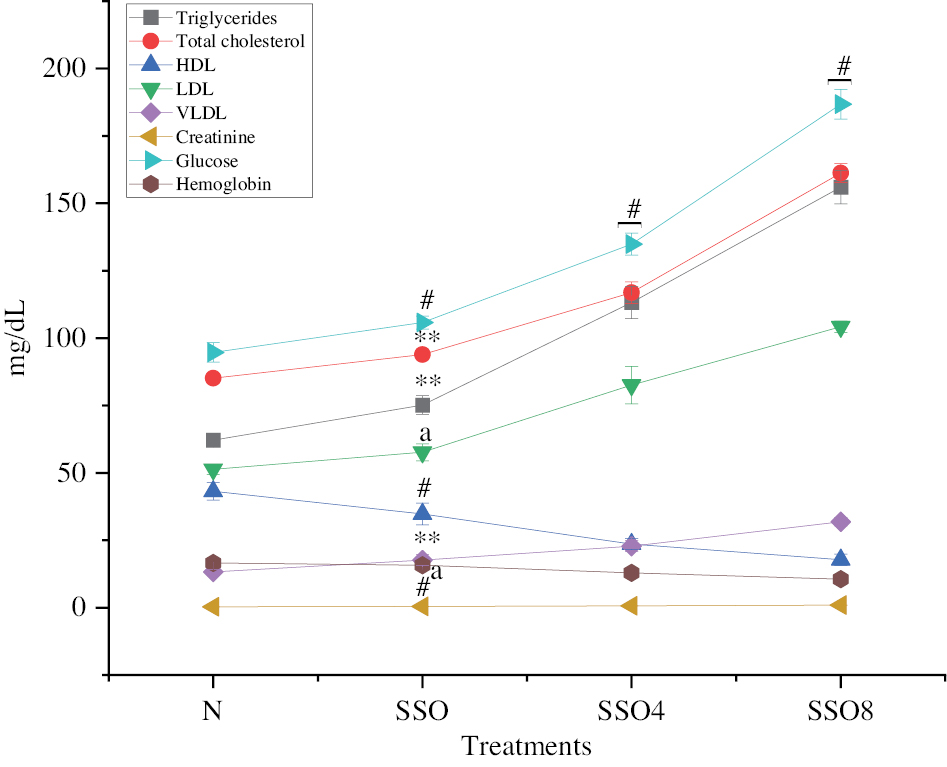
Figure 3 Liver enzymes in groups treated with normal fresh oil or thermo-oxidized sesame seed oil (SSO) under various conditions. The experimental conditions were as described in Figure 2. Oil samples were prepared by thermal oxidation at 180°C for 4 h or 8 h. Liver enzymes including ALT and AST were analyzed with an auto analyzer. The results are expressed as mean ± SD (n=6). #P<0.001, anon-significant values, as compared with the normal groups.
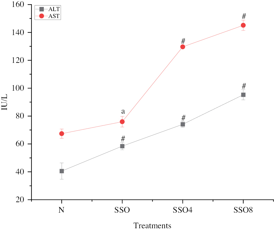
Figure 4 RBC counts in normal fresh oil and thermo-oxidized sesame seed oil (SSO) treated groups under various conditions. The experimental conditions were as described in Figure 2. Oil samples were prepared by thermal oxidation at 180°C for 4 h or 8 h. Blood was collected after the treatment, and RBC counts were analyzed with an autoanalyzer. The results are expressed as mean ± SD (n=6). #P<0.001, *P<0.05 as compared with the normal groups.
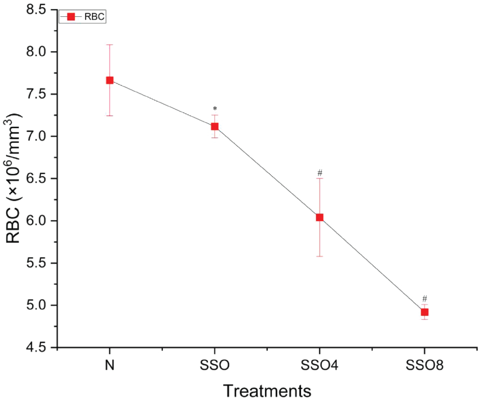
Figure 5 WBC and platelet counts in groups treated with normal fresh oil or thermo-oxidized sesame seed oil (SSO) under various conditions. The experimental conditions were as described in Figure 2. Oil samples were prepared by thermal oxidation at 180°C for 4 h or 8 h. Blood was collected after the treatment, and WBC and platelet counts were analyzed with an autoanalyzer. The results are expressed as mean ± SD (n=6). #P<0.001, *P<0.05, and anon-significant values, as compared with the normal groups.
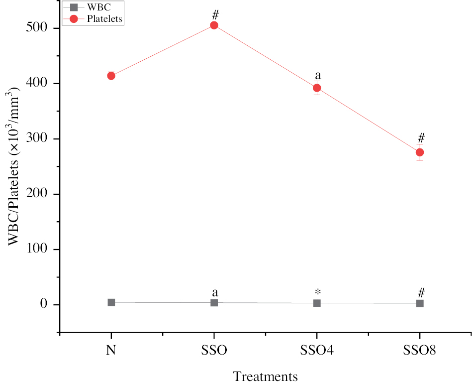
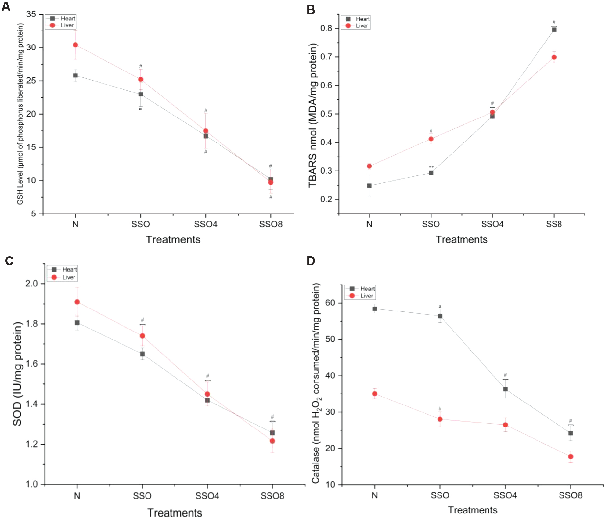
Figure 6 (A) Glutathione (GSH) levels in heart and liver tissue. We examined GSH levels in heart and liver tissues from rats treated with normal fresh oil or thermo-oxidized sesame seed oil (SSO) under various conditions. GSH levels decreased, thus indicating oxidative damage in the tissue. (B) Thiobarbituric acid reactive substance (TBARS) levels in heart and liver tissue. Similarly, we assessed TBARS levels in the same tissue samples. TBARS levels increased, thus further confirming oxidative damage due to SSO treatment. (C) Superoxide dismutase (SOD) levels in heart and liver tissue: We also measured SOD levels in the heart and liver tissue of rats exposed to SSO. Interestingly, SOD levels decreased, in agreement with oxidative damage. (D) Catalase levels in heart and liver tissue. Finally, we evaluated catalase levels in the same tissue samples. Catalase levels decreased, thus reinforcing the presence of oxidative damage caused by SSO treatment. The results are expressed as mean ± SD (n=6). #P<0.001, **P<0.01, *P<0.05, and a non-significant values, as compared with the normal groups.
The authors would like to apologize for this error and any inconvenience caused.
DOI of original article https://doi.org/10.15212/bioi-2024-0045